XB-IMG-116418
Xenbase Image ID: 116418
|
|
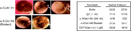
Figure 4. Injection of anti-XCdc14α/β antibodies into Xenopus embryos blocks cell division. One cell of 2-cell Xenopus embryos was injected with 10 nL of XB Buffer, purified rabbit IgG (final concentration ~1 μM), anti-XCdc14α/β antibodies (~200 nM final concentration), anti-XCdc14α/β antibodies pre-incubated with the immunizing antigen ("Blocked"), or GST-hCdc14A (1 μM). Left: examples of embryos injected with α-XCdc14α/β antibodies (top) or pre-blocked α-XCdc14α/β antibodies. Right: summary of two independent experiments. Embryos were analyzed at the 32-cell stage, and only embryos with an obvious large blastomere phenotype were considered abnormal. Image published in: Kaiser BK et al. (2004) Image downloaded from an Open Access article in PubMed Central. Copyright © 2004 Kaiser et al; licensee BioMed Central Ltd. This is an Open Access article: verbatim copying and redistribution of this article are permitted in all media for any purpose, provided this notice is preserved along with the article's original URL. Larger Image Printer Friendly View |