Click here to close
Hello! We notice that you are using Internet Explorer, which is not supported by Xenbase and may cause the site to display incorrectly.
We suggest using a current version of Chrome,
FireFox, or Safari.
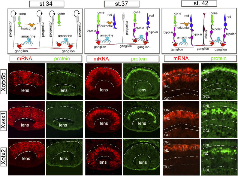
Figure 2. The Translation of the Xenopus Homeobox Xotx5b, Xvsx1, and Xotx2 mRNAs Parallels the Generation of Photoreceptors and Bipolar CellsIn situ hybridization of Xotx2, Xotx5b, and Xvsx1 mRNAs (Fast Red detection) compared to immunostaining of the corresponding proteins (green detection) on serial 10-μm sections of embryonic retinas at st. 34 (mid-neurogenesis), st. 37 (late-neurogenesis), and st. 42 (mature embryonic retina). Schematics show the retinal cell types present at the corresponding times of analysis (see also Figure S2). Dashed lines border the entire thickness of neural retinas (st. 34â37), or indicate the boundaries between different cell layers (st. 42, magnification of central retinal aspect); GCL: ganglion cell layer, INL: inner nuclear layer, ONL: outer nuclear layer.