XB-IMG-116866
Xenbase Image ID: 116866
|
|
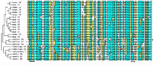
Figure 6. Alignment of the duplicated FN type III domains oftenascin-W. The third FN type III domain of Tetraodon nigroviridis tenascin-W has been duplicated one or more times in other tensacin-Ws. Alignment reveals the conservation of sequences within these domains, including putative integrin binding motifs near the N-terminus of the domain (underlined). The region where an integrin-binding RGD sequence in an exposed loop is found in chicken tenascin-C is indicated by asterisks. Several tenascin-Ws have a potentially active KGD motif in this region. At the left is a rooted phylogenetic tree generated by SATCHMO. This analysis indicates that many of the domain duplications took place after the divergence of primate and rodent lineages. Identical amino acids are shaded blue, while similar amino acids are boxed in yellow. Image published in: Tucker RP et al. (2006) Copyright © 2006 Tucker et al; licensee BioMed Central Ltd. This image is reproduced with permission of the journal and the copyright holder. This is an open-access article distributed under the terms of the Creative Commons Attribution license Larger Image Printer Friendly View |