XB-IMG-118097
Xenbase Image ID: 118097

|
|
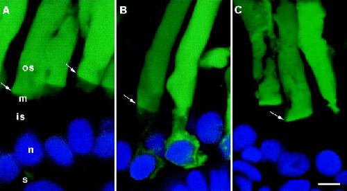
Figure 7. Presentation of the targeting signal affects the efficiency of ROS localization. Confocal micrographs of cells expressing (A) mGFP-CT9; (B) mGFP-CT25; and (C) mGFP-CT44C322/323S. Varying the lengths of the rhodopsin peptides fused to GFP affected the distribution patterns. mGFP-CT9 and mGFP-CT44C322/323, but not mGFP-CT25, were significantly more efficient that mGFP (see Fig. 4 E) in targeting to the ROS. Mitochondria were also labeled to varying degrees by the myristoylated fusion proteins. Arrows indicate the inner/outer segment junction. GFP (green) and Hoescht 33342 (blue). os, outer segment; is, inner segment; n, nucleus; m, mitochondria; and s, synaptic terminal. Bar, 5 μm. Image published in: Tam BM et al. (2000) © 2000 The Rockefeller University Press. Creative Commons Attribution-NonCommercial-ShareAlike license Larger Image Printer Friendly View |
