XB-IMG-124629
Xenbase Image ID: 124629
|
|
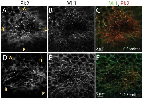
Figure 1. VANGL1 and PRICKLE2 set up PCP in the mouse ventral node.(A–F) Localization of VL1 and PK2 proteins in the ventral node: PRICKLE2 is expressed in the node of 0 somite embryos (A, C red) prior to VANGL1, which is detected in node cells (E, F, green) of 1–2 somite embryos. VL1 and PK2 co-localize in node cells and form crescents pointed toward the anterior (F, yellow). Motile cilia can be visualized above the plane of VL1 or PK2 localization (not shown). Yellow letters mark anterior (A), posterior (P), Left (L) and right (R). As nodes were imaged from the ventral side, left side of the embryo is on the right side of each panel and right side of the embryo is on the left side of each panel. Image published in: Antic D et al. (2010) Antic et al. This image is reproduced with permission of the journal and the copyright holder. This is an open-access article distributed under the terms of the Creative Commons Attribution license Larger Image Printer Friendly View |