XB-IMG-124729
Xenbase Image ID: 124729
|
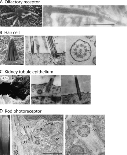
Figure 1. Ultrastructures of a sampling of primary cilia. (A) Olfactory receptor cilia
contain the molecular machinery of odorant transduction. Defects in ciliary
genes lead to anosmia (Kulaga et al.,
2004). (Left) Scanning EM of an embryonic rat olfactory receptor
dendritic knob with multiple primary cilia (reproduced from Menco, 1997 with permission of Oxford
University Press). (Right) Longitudinal section through an olfactory cilium
(reproduced from Menco et al., 1997
[fig. 6] with permission from
Springer Science and Business Media). Bar, 1 µm. (B) Hair cells are
mechanoreceptors. They generally have only one true cilium, the kinocilium,
the longest structure found at the back of the ciliary bundle. Defects in
ciliary genes lead to reduced hearing and deafness, such as in
Usher’s syndrome. (Left) Scanning EM of stereocilia from a frog
saccule (reproduced from Vollrath et al.,
2007 with permission). Bar, 1 µm. (Middle and right)
Sections through the kinocilia of teleost fish (reproduced from Flock and Duvall, 1965 with
permission). (Middle) Longitudinal section through the kinocilium base. Bar,
0.5 µm. (Right) Cross section showing a 9 + 2 axonemal
structure. Bar, 0.1 µm. (C) Kidney tubule epithelial cells possess
single cilia, whose function remains unknown, but it has been proposed that
they serve some sensory role, such as the detection of fluid flow. Mutations
in cilia genes lead to polycystic kidney disease and loss of renal function.
(Left) Scanning EM of mouse kidney tubule epithelial cells showing several
cilia projecting into the lumen (Pazour et
al., 2000). (Middle and right) Longitudinal sections through the
base of the cilia showing the basal body and centriole (reproduced from
Ganote et al., 1968 with
permission). (D) Photoreceptors in the retina are modified cilia that
transduce light into visual signals. The OSs contain opsin molecules that
absorb photons and pass the light signal down a transduction cascade that
leads to channel closure in the plasma membrane. Mutations in ciliary genes
lead to retinal degeneration and blindness, such as in retinitis pigmentosa.
(Left) Scanning EM of a frog rod. (Middle) Longitudinal section through the
CC showing the basal body and the associated centriole. Note the lamellar
discs (D) of the OS and the mitochondria (M) of the IS compartments. Bar, 1
µm. (Right) Cross section of the CC just distal to the basal body.
Bar, 0.5 µm. Images reproduced from Peters et al. (1983) with permission. Image published in: Calvert PD et al. (2010) © 2010 Calvert et al. This image is reproduced with permission of the journal and the copyright holder. This is an open-access article distributed under the terms of the Creative Commons Attribution-NonCommercial-ShareAlike license Larger Image Printer Friendly View |