XB-IMG-125193
Xenbase Image ID: 125193
|
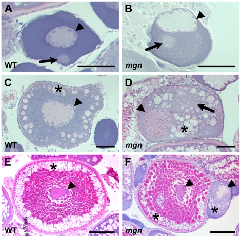
Figure 2. The mgn mutation causes defects during oogenesis.Sections of wild-type and mutant oocytes stained with hematoxylin (purple) and eosin (pink). The cytoplasm of stage I zebrafish oocytes is strongly basophilic, resulting in strong purple staining, while the mitochondria-rich Balbiani body is slightly acidophilic and stains pale pink with eosin. In wild-type mid stage I oocytes (A), the nucleus is localized at the center of the oocyte and the Balbiani body is near the future vegetal cortex. In mgn mutant stage I oocytes (B), the nucleus is asymmetrically localized and the Balbiani body, which frequently remains close to the nucleus, is surrounded by a region that is lightly stained with eosin. Wild-type stage II oocytes (C) have a central nucleus surrounded by cortical granules (CG). In stage II mgn mutant oocytes (D), the nucleus is mislocalized and CG accumulate opposite to the nucleus, in and around a faint eosin-stained area (arrow). During stage III, wild-type oocytes (E) accumulate yolk in the center of the oocyte and CG localize uniformly around the cortex, whereas stage III mgn mutant oocytes (F) display an uneven distribution of CG at the cortex. Also note stage II oocyte at right in (F). Arrowheads indicate oocyte nuclei; arrows indicate Balbiani body in (A,B); asterisks indicate CG. Scale bars = 50 microns (A,B), 100 microns (C,D), and 200 microns (E,F). Image published in: Gupta T et al. (2010) Gupta et al. This image is reproduced with permission of the journal and the copyright holder. This is an open-access article distributed under the terms of the Creative Commons Attribution license Larger Image Printer Friendly View |