XB-IMG-127277
Xenbase Image ID: 127277
|
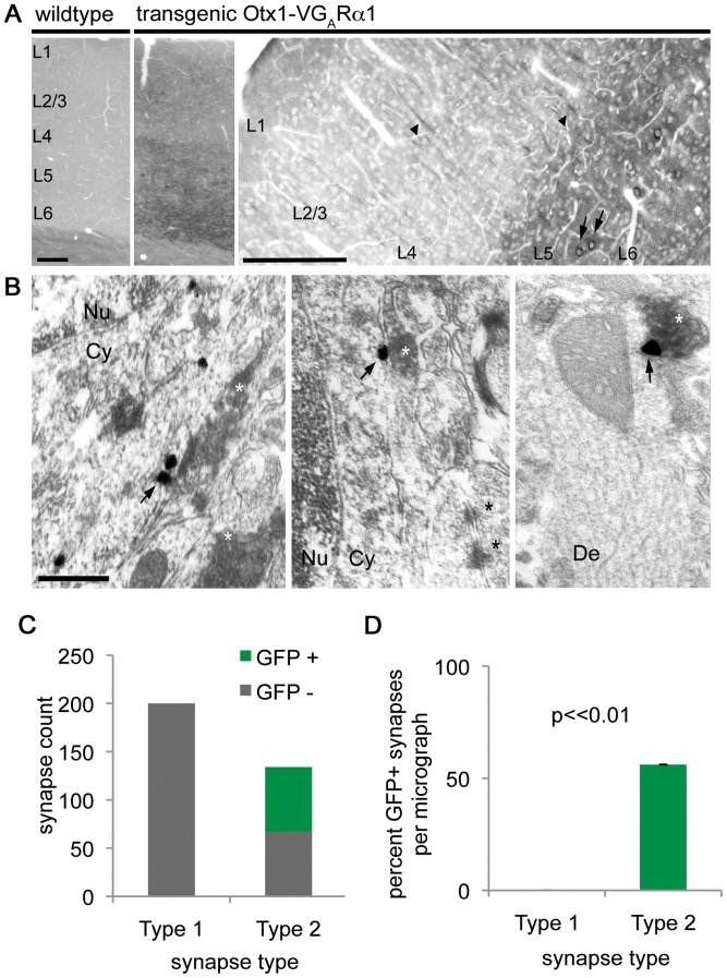
Figure 3. Venus-GABAARα1 localizes specifically to inhibitory synapses.(A) Light microscopy of fixed saggital sections from wild type and Otx1-VGABAARα1 transgenic mice treated with anti-GFP antibody and revealed with the DAB procedure. Transgenic, but not wild type mice express the fusion protein in layer 5/6 cortical pyramidal neurons. The fusion protein localizes to cell bodies (arrows) and processes (arrowheads) in cortex. Scale bars: 200 µm. (B) Immuno-electron microscopy shows VGABAARα1 expression (arrows) exclusively at inhibitory synapses by silver-intensified immunogold labeling (SIG). Inhibitory terminals immunoreactive for GAD65/67 are revealed with the DAB procedure (white asterisks). Asymmetric synapses (black asterisks) are immunonegative for both GAD and VGABAARα1. Scale: 500 nm. Cy: cytoplasm. Nu: nucleus. De: dendrite. V: Venus. GAR: GABAA receptor. (C) Within a total cortical area of 614.6 square microns 67 of the 134 inhibitory (symmetric) synapses were labeled by VGABAARα1, whereas none of the 200 excitatory (asymmetric) synapses were immunopositive for the fusion protein. (D) An average of 54% of inhibitory synapses were immunopositive for VGABAARα1, compared to 0% of the excitatory synapses. The data are presented as average ± SEM (t test). Image published in: Heller EA et al. (2012) Heller et al. Creative Commons Attribution license Larger Image Printer Friendly View |