XB-IMG-127283
Xenbase Image ID: 127283
|
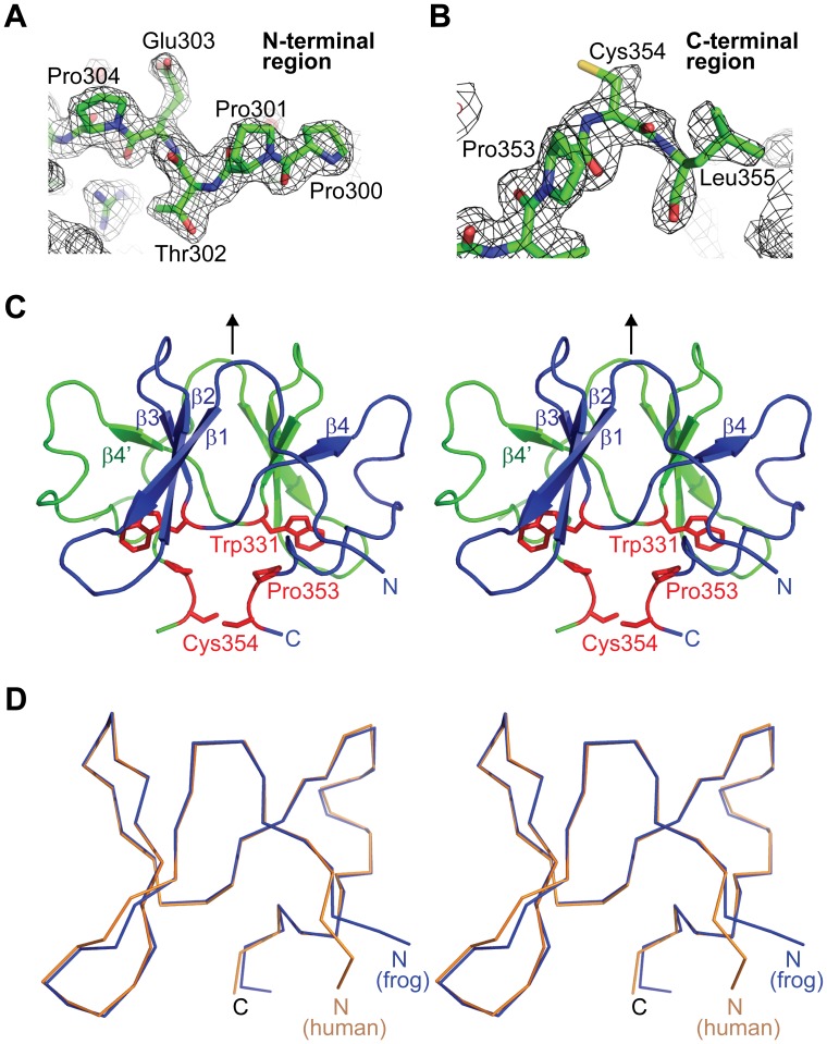
Figure 3. Crystal structure of the frog DGCR8 dimerization domain.(A–B) 2Fo-Fc electron density maps, contoured at 1σ level, of the N- and C-terminal regions of the frog dimerization domain, respectively. (C) Wall-eyed stereo diagram of the crystal structure of frog dimerization domain. The dimer subunits are colored green and blue. Secondary structures from the green subunit are denoted with a prime. The crystallographic two-fold axis relating the two subunits is indicated by the arrow. Residues known to be important for heme binding are highlighted in red. (D) Superimposition of human (orange) and frog (blue) dimerization domain Cα traces shown in stereo. Image published in: Senturia R et al. (2012) Senturia et al. This image is reproduced with permission of the journal and the copyright holder. This is an open-access article distributed under the terms of the Creative Commons Attribution license Larger Image Printer Friendly View |