XB-IMG-130644
Xenbase Image ID: 130644

|
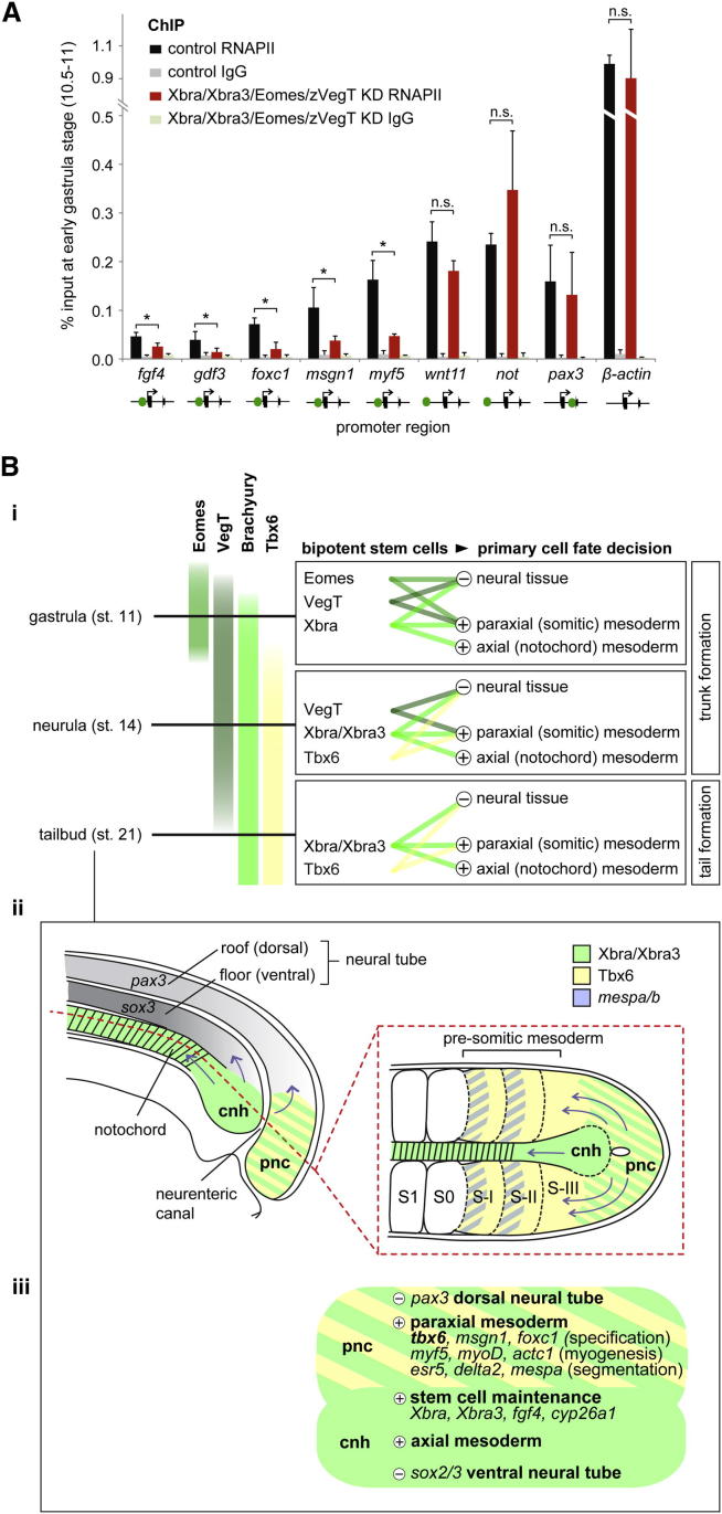
Figure 7. T-box TF-Dependent Recruitment of RNAPII and Model for the Way in which Stage-Dependent Combinations of T-box TFs Define and Instruct Bipotential Stem Cells to Be Recruited to Neural and Mesodermal Tissues and Prime Mesoderm for Differentiation(A) RNAPII deposition at promoters of mesodermal (fgf4, gdf3, foxc1, msgn1, myf5, wnt11, not), neural (pax3), and house-keeping (β-actin) genes in control and T-box TF-depleted embryos at early gastrula stage (10.5–11) determined by ChIP-qPCR. Proximal or distal (upstream or intronic) binding of T-box TFs to indicated gene promoter is symbolized with green dot. The error bars represent SD of biological triplicates. Two-tailed Student’s t test: ∗p < 0.1; n.s., not significant (p ≥ 0.1). IgG, immunoglobulin G.(B) Model: (i) The different spatial and temporal patterns of T-box TFs cause neuromesodermal stem cells to be defined by Eomes, VegT, and Xbra during gastrulation; VegT, Xbra, Xbra3, and Tbx6 during neurulation; and Xbra, Xbra3, and Tbx6 during tail bud stages. These combinations of T-box TFs also ensure that the correct ratio of mesodermal over neural tissue is formed during trunk and tail formation by activating mesoderm-specifying genes and repressing neurogenic genes. The development of axial (notochord) mesoderm depends mainly on Xbra/Xbra3, due to their exclusive expression among these T-box TFs in the chordoneural hinge and developing notochord. Other mesodermal derivatives, such as heart, may similarly depend on combinations of T-box TFs (omitted from model). (ii) Schematic diagram of a sagittal section and a horizontal section (red dashed line) through the posterior region of an early tail bud embryo illustrating the expression of Xbra/Xbra3, Tbx6, and mespa/b and the recruitment of mesodermal and neural cells (blue arrows) from the stem niche (chordoneural hinge and posterior wall of the neurenteric canal). Most cells of the chordoneural hinge give rise to the notochord and the ventrolateral horns of the neural tube, whereas cells in the posterior wall of the neurenteric canal contribute to paraxial (presomitic) mesoderm and the dorsal roof of the neural tube. cnh, chordoneural hinge; pnc, posterior wall of neurenteric canal; S1, first somite; S0, newly forming somite; S-I/II/III, presomitic mesoderm. (iii) Genetic regulatory inputs of T-box TFs in early tail bud embryos with several functional nodes being active in different domains (cnh, pnc) of the tail bud: stem cell maintenance; specification of somitic mesoderm; myogenic differentiation; patterning of presomitic mesoderm; notochord formation; and protection from neuralization. Image published in: Gentsch GE et al. (2013) © 2013 The Authors. Creative Commons Attribution license Larger Image Printer Friendly View |
