XB-IMG-130816
Xenbase Image ID: 130816
|
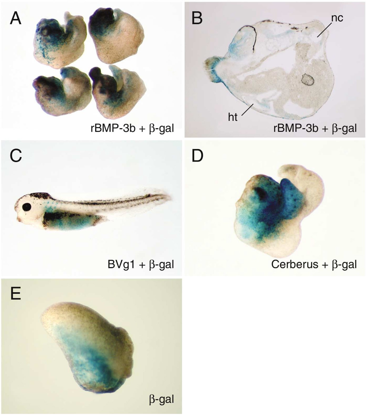
Fig. 4. BMP-3b triggers head formation in an intrinsic developmental pathway. To examine head formation by BMP-3b, we performed lineage tracing and
rescue of embryos exposed to UV. (A) Injection of rBMP-3b mRNA (1 ng) rescued anterior structure of UV-ventralized embryos. Cells expressing BMP-3b
were localized in head region (60%, n 15). (B) Section of an embryo shown in (A). Cells were labeled in notochord (nt), heart (ht), optic cup, and adjacent
to cement gland, suggesting that cells expressing BMP-3b differentiated into âhead-forming cellsâ and organized head structures. (C) Cells expressing BVg1
(50 pg), an organizer inducer, were localized in endoderm, suggesting that they induced head-forming cells (50%, n 14; Thomsen and Melton, 1993). (D)
Injection of cerberus (1 ng) led to formation of smaller head than that with BMP-3b (60%, n 25; Bouwmeester et al., 1996). (I) Control embryo. Image published in: Hino J et al. (2003) Copyright © 2003. Image reproduced with permission of the Publisher, Elsevier B. V. Larger Image Printer Friendly View |