XB-IMG-131059
Xenbase Image ID: 131059
|
|
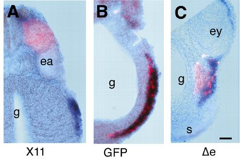
Fig. 3. Localisation of host and donor CNC cells at stage 28. (A) Transverse section showing a Xcad-11-expressing non-migrating transplant (pink), and migrating host CNC cells (blue). (B) Mixture of host (blue) and donor (pink) CNC cells in GFP-RNA injected control embryos. (C) Transverse section showing most of the δeXcad-11-expressing, migrating donor CNC cells (pink) separated from the host CNC cells (blue). Pink, immunostaining of Myc-tagged GFP; blue, twist in situ hybridisation. ea, ear vesicle; ey, eye anlage; g, gut. Scale bar: 50 μm. Image published in: Borchers A et al. (2001) Copyright © 2001. Image reproduced with permission of the publisher and the copyright holder. This is an Open Access article distributed under the terms of the Creative Commons Attribution License. Larger Image Printer Friendly View |