XB-IMG-131631
Xenbase Image ID: 131631
|
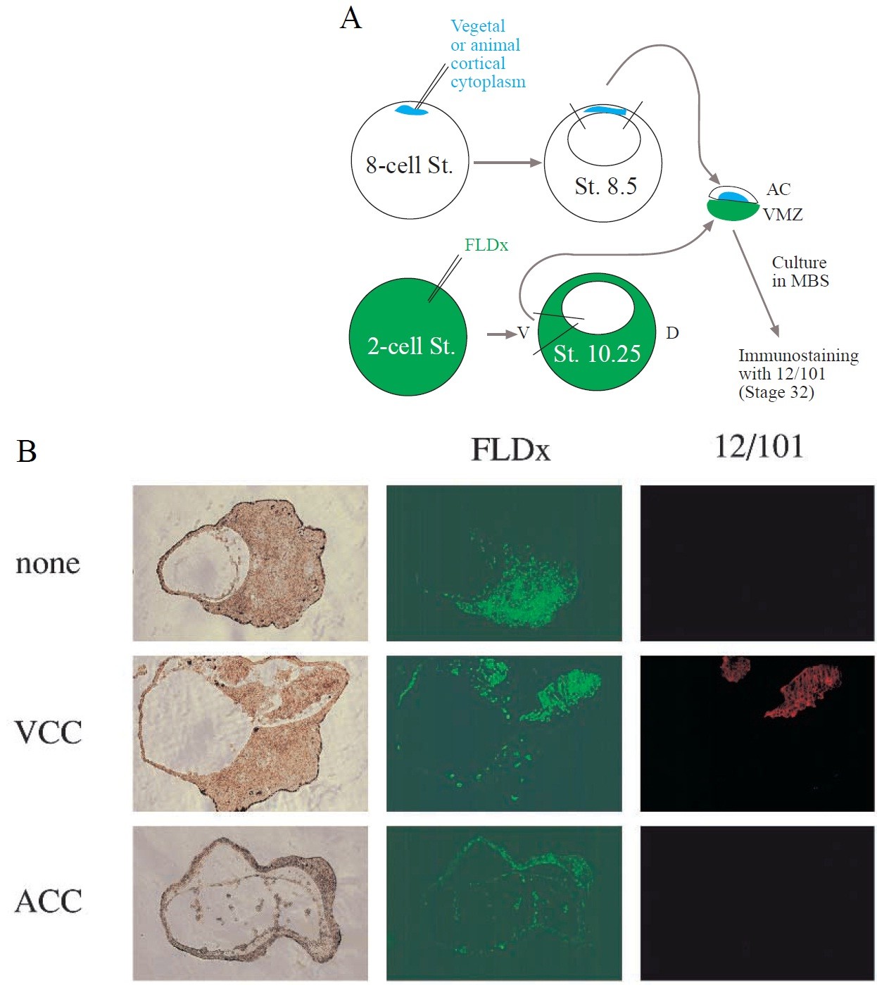
Fig. 3. Animal caps injected with vegetal cortical cytoplasm secrete a dorsalising signal. (A) Diagrammatic
representation of the experiment. Ventral marginal zone explants (composed mostly of ventral mesodermal cells)
were taken from embryos (stage 10.25) previously injected with the lineage tracer FLDx. The explants were
immediately combined with blastula animal caps (stage 8.5) derived from embryos previously injected with
animal or vegetal cortical cytoplasm (ACC, VCC). The conjugates were cultured until their mesodermal
component reached the equivalent of stage 32 and they were then immunostained with the muscle antibody
12/101. (B) Photographs of sections through conjugates composed of a ventral marginal zone explant labelled
with fluoresceine lysinated dextran (FLDx) combined with an animal cap derived either from uninjected embryos
(none), from embryos injected with vegetal cortical cytoplasm (VCC), or from embryos injected with animal
cortical cytoplasm (ACC). Left panels, bright-field images; middle panels, position of FLDx-labelled cells; right
panels, 12/101 staining. Muscle-specific staining is seen only in the progeny of the FLDx-labelled marginal zone
cells which were conjugated to the VCC-injected animal cap. Image published in: Darras S et al. (1997) Copyright © 1997. Image reproduced with permission of the publisher and the copyright holder. This is an Open Access article distributed under the terms of the Creative Commons Attribution License. Larger Image Printer Friendly View |