XB-IMG-132244
Xenbase Image ID: 132244
|
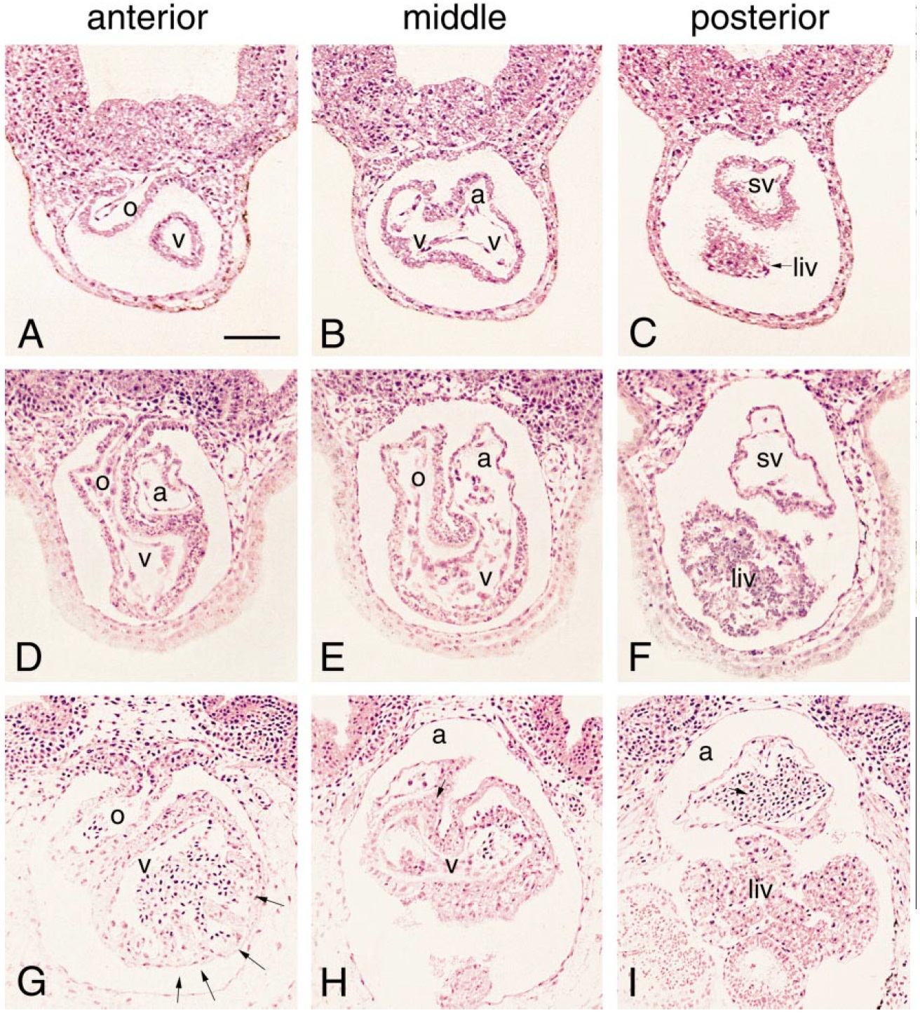
FIG. 8. The acquisition of distinct chamber morphologies is clearly resolved in transverse sections from methacrylate-embedded embryos.
(A–C, D–F, and G–I) Sections through the anterior, middle, and posterior regions of the heart tube at stages 35, 39, and 42, respectively. At
the onset of looping (stage35), the myocardial walls of the outflow tract (o), ventricular (v), atrial (a), and posterior sinus (sv) regions are of
equal thickness. By stage 39, differential thickening is evident in the outflow tract and ventricular region. At stage 42, trabeculae (arrows)
have formed in the lateroventral wall of the ventricular myocardium. (Blood cells are evident throughout the heart at this stage.) Bar
represents 100 mm. Image published in: Mohun TJ et al. (2000) Copyright © 2000. Image reproduced with permission of the Publisher, Elsevier B. V. Larger Image Printer Friendly View |