XB-IMG-133938
Xenbase Image ID: 133938
|
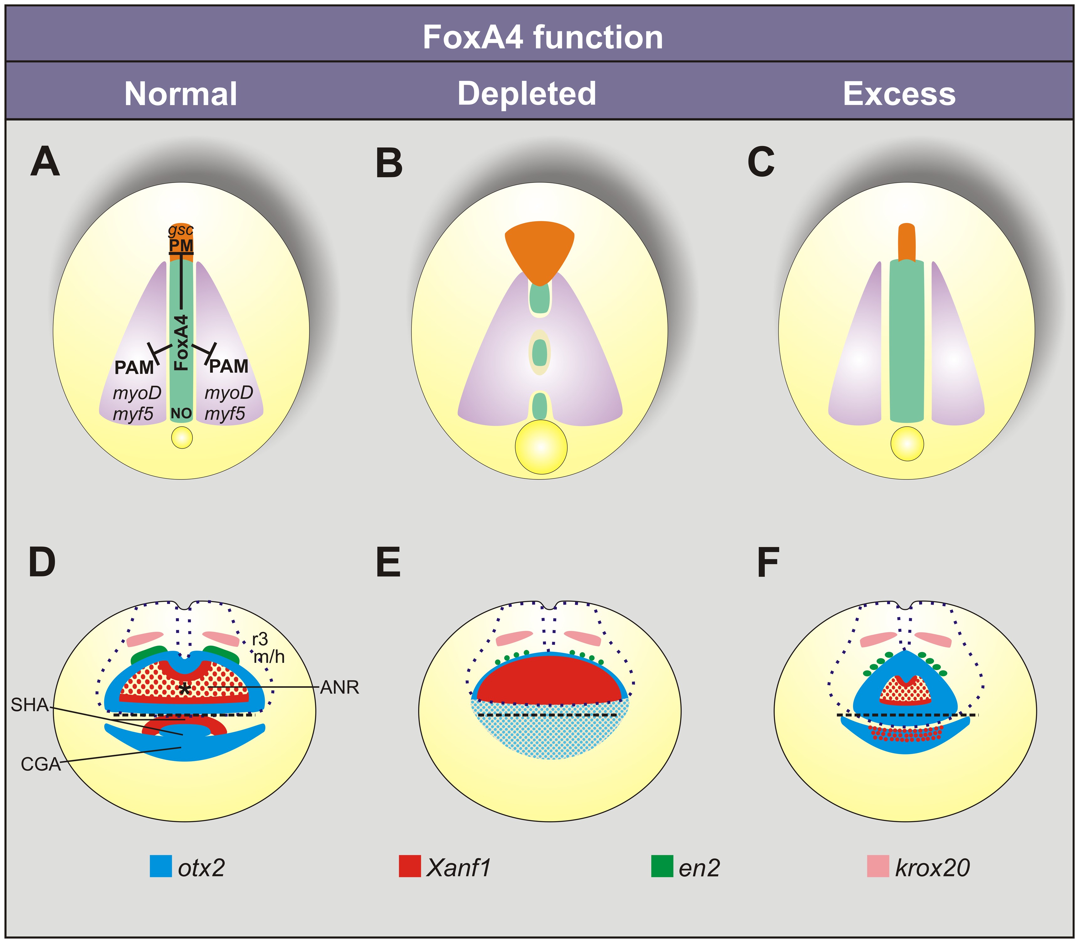
Figure 10. Summary of the phenotypes after manipulating foxA4 function in Xenopus embryos.
The diagrams show the phenotypes in embryos with normal (A, D), depleted (B, E), or excess levels (C, F) of foxA4 function, as revealed by the expression patterns of bra, chd, myf5/myoD, gsc, frzb1, otx2, Xanf1, krox20 and en2 at neural plate stage. (A–C) In DML development, foxA4 is necessary to restrict the paraxial mesodermal (PAM) and the prechordal mesodermal (PM) fates, allowing notochord (NO) development. (D–F) Controlled levels of foxA4 function are necessary for the correct specification and segregation of the anterior neural and ectodermal anlagen. ANR, anterior neural ridge; SHA, stomodeal-adenohypophyseal anlage; CGA, cement gland anlage; r3, third rhombomere; m/h, midbrain/hindbrain boundary. The asterisk in D marks the expression hole left by otx2 in the ANR, which is occupied by Xanf1. The dotted blue line (D–F) represents the contour of the neural plate. The dashed black line (D–F) represents the anterior boundary of the neural ectoderm. (A–C) Dorsal views. (D–F) Anterior views. See text for details.
doi:10.1371/journal.pone.0110559.g010 Image published in: Murgan S et al. (2014) Image reproduced on Xenbase with permission of the publisher and the copyright holder. This image is reproduced with permission of the journal and the copyright holder. This is an open-access article distributed under the terms of the Creative Commons Attribution license Larger Image Printer Friendly View |