XB-IMG-134394
Xenbase Image ID: 134394

|
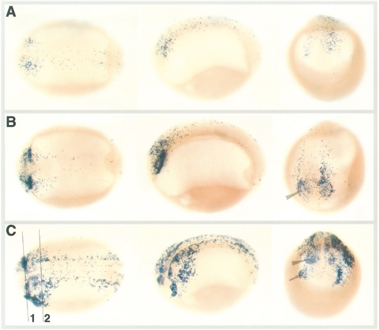
FIG. 3. Programmed cell death as detected by whole-mount TUNEL staining at the neural fold stage. A, B, and C, stage 17. Dorsal, lateral,
and end-on anterior views are shown for each embryo (left to right). (A) TUNEL staining was detected in the anteriormost portion of the
embryo with a light scattering of staining along the length of the neural fold. (B) Strong TUNEL staining in the anteriormost portion of the
embryo, corresponding to the optic placodes (arrowhead). Staining extends from these placodes along the neural fold. (C) Strongest staining
was in the anterior embryo with patches of TUNEL staining corresponding to the optic (arrowhead), and olfactory (arrow) placodes, and
developing brain. Dying cells were found in defined stripes on each side of the dorsal midline corresponding to the primary sensory neurons.
A scattering of dying cells was detected throughout the neural fold. Lines 1 and 2 mark the approximate location of transverse sections
shown in Figs. 7B and 7C, respectively. The cell death patterns are representative of the staining observed following staining of 33 embryos
of which 64% were TUNEL positive. Embryos with more than 5 TUNEL-stained nuclei were considered TUNEL positive. Image published in: Hensey C and Gautier J (1998) Copyright © 1998. Image reproduced with permission of the Publisher, Elsevier B. V. Larger Image Printer Friendly View |
