XB-IMG-134398
Xenbase Image ID: 134398
|
|
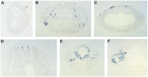
FIG. 7. Sections of TUNEL-stained embryos from stages 13 to 35. (A) Stage 13, arrowhead indicates TUNEL staining in the presumptive
neuroectoderm. (B) Stage 17, transverse section (1) indicated in Fig. 3C, shows TUNEL staining in circular patches which correspond to the
developing brain and sensory placodes. (C) Stage 17, a transverse section (2) indicated in Fig. 3C, shows TUNEL staining in the region of
the neural crest. (D) Stage 26, a transverse section indicated in Fig. 5A, TUNEL staining was detected in both sides of the spinal cord giving
rise to two bilaterally symmetrical dorsal stripes, Fig. 5A (arrowheads). (E) Stage 31/32, a transverse section through the head as indicated
in Fig. 6B shows TUNEL staining in the brain and optic stalk. Most intense staining is evident in the ventral diencephlon. (F) Stage 29/30,
a sagital section of the embryo indicated in Fig. 6A, shows TUNEL staining in the mesencephalon, diencephalon, and telencephalon regions
of the brain. Image published in: Hensey C and Gautier J (1998) Copyright © 1998. Image reproduced with permission of the Publisher, Elsevier B. V. Larger Image Printer Friendly View |