XB-IMG-134652
Xenbase Image ID: 134652
|
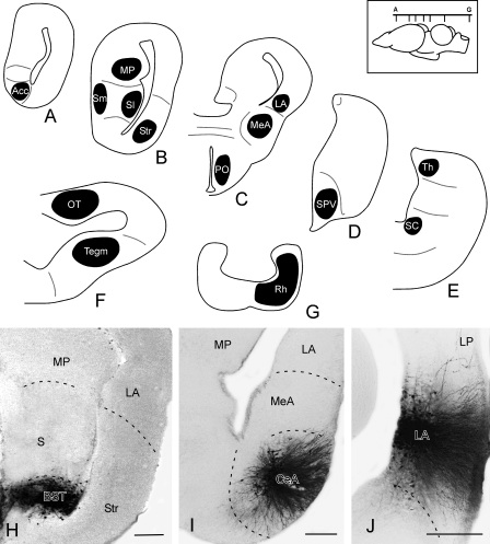
Figure 10. Schematic drawings of transverse sections through the brain of X. laevis illustrating the representative injection sites used in the confirmation experiments. The levels of the selected transverse sections (A–G) are indicated in the upper right scheme of the brain. H–J: Photomicrographs showing the examples of injection sites in the bed nucleus of the stria terminalis (H), central amygdala (I), and lateral amygdala (J). Scale bars = 100 μm. Image published in: Domínguez L et al. (2014) Copyright © 2014. Image reproduced with permission of the Publisher. Larger Image Printer Friendly View |