XB-IMG-134653
Xenbase Image ID: 134653
|
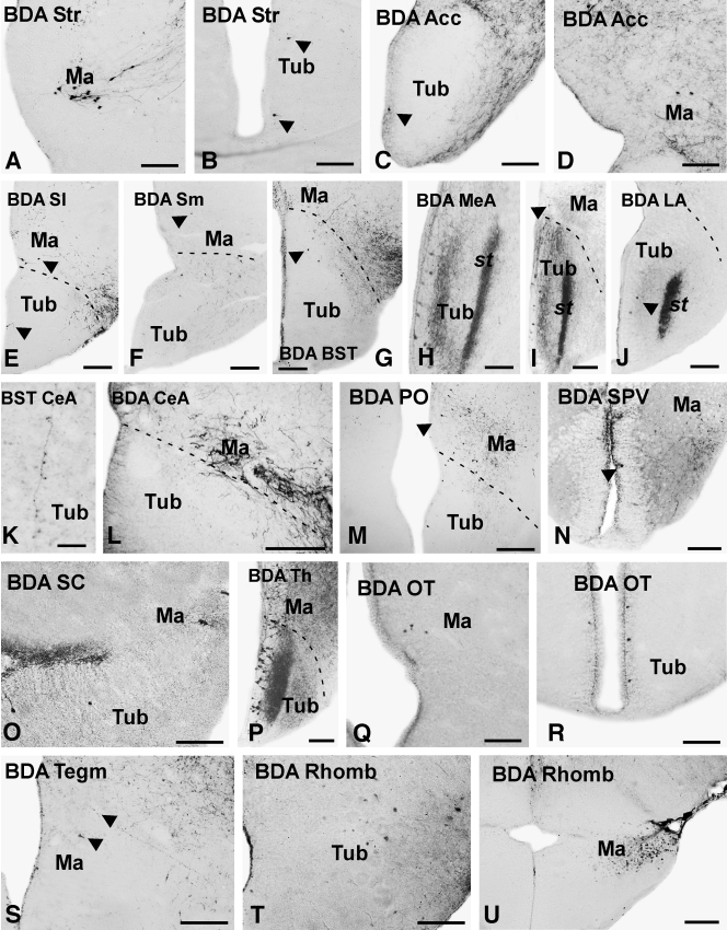
Figure 11. Photomicrographs of transverse sections through the brain of X. laevis showing retrogradely labeled cells and anterogradely labeled fibers in the tuberal and mammillary regions after BDA injections carried out in the different brain centers indicated in each photograph. The arrowheads in (B,C,E–G,I,J,M,N,S) point to labeled cells and fibers. Scale bars = 200 μm in B,C,E–J,L–N,P,R,U; 100 μm in A,D,O,Q,S,T; 50 μm in K. Image published in: Domínguez L et al. (2014) Copyright © 2014. Image reproduced with permission of the Publisher. Larger Image Printer Friendly View |