Click here to close Hello! We notice that you are using Internet Explorer, which is not supported by Xenbase and may cause the site to display incorrectly. We suggest using a current version of Chrome, FireFox, or Safari.
XB-IMG-134810

Xenbase Image ID: 134810
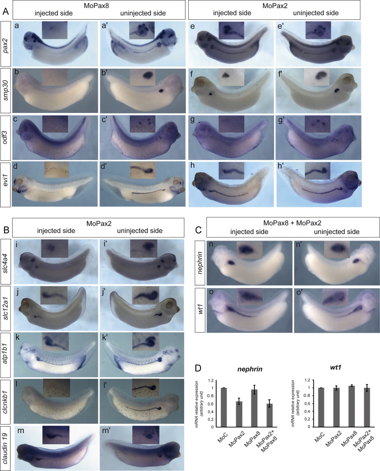

Fig. 2. Pax 8 and Pax2 loss of function differentially affects tubule but not glomus marker genes expression at late tailbud stage. (A, B) Whole mount in situ hybridization for tubule marker genes. Embryos were injected with MoPax8 (ad) or MoPax2 (em) at the 4-cell stage, in the equatorial region of both left blastomeres. They were cultured until the tailbud stage 3335, fixed and analyzed by in situ hybridization with specific probes for the indicated genes. For each embryo, injected (am) and uninjected (am) sides are shown. Pax8 depletion prevents expression of marker genes all along the entire tubule expression domain. In Pax2 morphants, gene expression in the proximal part of the tubule, including nephrostomes, is affected. Expression in the distal tubule is not modified by the MoPax2 except for the terminal differentiation marker clcnkb1 whose expression is completely inhibited. Insets show higher magnification of the anterior pronephric territory. (C) Whole mount in situ hybridization for the glomus marker genes wt1 and nephrin. Embryos were injected with a mixture of MoPax8 and MoPax2 in the equatorial region of both left blastomeres at the 4-cell stage and cultured until the tailbud stage 33. Injected sides are shown in (n, o), uninjected sides in (n, o). Insets show higher magnification of the anterior pronephric territory. (D) RT-qPCR analysis of wt1 and nephrin. Embryos were injected with the indicated Mo at the 4-cell stage in all four blastomeres. They were cultured until the late tailbud stage 33 and processed for RT-qPCR analysis. Average values from three independent experiments. Neither MoPax2 nor MoPax8 injected alone or together significantly affects wt1 and nephrin expression compared to the control (MoC).

Image published in: Buisson I et al. (2015)

Copyright © 2015. Image reproduced with permission of the Publisher, Elsevier B. V.

GeneSynonymsSpeciesStage(s)Tissue
pax2.LLOC108697493, pax-2, pax2-a, pax2-b, XPax-2, XPax2X. laevisSometime during NF stage 33 and 34 to NF stage 35 and 36pronephric tubule
kidney
pronephric kidney
optic stalk
hindbrain
spinal cord
post-anal gut
pronephric duct
pronephric nephrostome
rgn.Lregucalcin, smp30, xsmp-30, xsmp30X. laevisSometime during NF stage 33 and 34 to NF stage 35 and 36kidney
glomus
pronephric kidney
cimap1a.Lodf3X. laevisSometime during NF stage 33 and 34 to NF stage 35 and 36pronephric nephrostome
pronephric kidney
kidney
mecom.Lecotropic viral integration site 1, evi-1, evi1, mecom-a, mecom-b, xEvi-1X. laevisSometime during NF stage 33 and 34 to NF stage 35 and 36pronephric tubule
kidney
pronephric duct
slc4a4.LhhNMC, hnbc1, nbc1, nbc2, nbce1, pNBC, slc4a5, XNBC1X. laevisSometime during NF stage 33 and 34 to NF stage 35 and 36pronephric kidney
glomus
pronephric nephrostome
kidney
slc12a1.LNa-K-2Cl cotransporter, NKCC-2, NKCC2, XNKCC2X. laevisSometime during NF stage 33 and 34 to NF stage 35 and 36pronephric kidney
glomus
pronephric duct
kidney
atp1b1.Satp1b1a, Beta1-Na/K-ATPase, Na+-K+-ATPase, β1-NaK-ATPaseX. laevisSometime during NF stage 33 and 34 to NF stage 35 and 36kidney
pronephric kidney
pronephric duct
glomus
post-anal gut
eye
early distal tubule
distal tubule
late distal tubule
proximal tubule
late proximal tubule
early proximal tubule
primary heart field
secondary heart field
forebrain
cement gland
clcnkb.LClC-k, clc-kb, clckb, clcnka, clk-k2, xClC-kX. laevisSometime during NF stage 33 and 34 to NF stage 35 and 36pronephric duct
early distal tubule
distal tubule
late distal tubule
early proximal tubule
late proximal tubule
proximal tubule
cldn19.Sclaudin-19X. laevisSometime during NF stage 33 and 34 to NF stage 35 and 36pronephric duct
glomus
nphs1.SnephrinX. laevisSometime during NF stage 33 and 34 to NF stage 35 and 36kidney
glomus
pronephric kidney
wt1.Swt-1, wt1-a, wt1-b, Xwt-1, xwt1X. laevisSometime during NF stage 33 and 34 to NF stage 35 and 36glomus
pronephric kidney
pronephric duct
dorsal lateral plate mesoderm

Image source: Published

Experiment + Assay Source Phenotypes and Disease
Xla Wt + pax2 MO + NF33/34-35/36 (in situ hybridization) Fig. 2 B, i-m
Expression Phenotype
decreased amount atp1b1.L expression in pronephric tubule
absent clcnkb.L expression in pronephric duct
absent clcnkb.L expression in pronephric kidney
decreased amount cldn19.L expression in proximal tubule
decreased amount slc12a1.L expression in proximal tubule
decreased amount slc4a4.L expression in proximal tubule
Xla Wt + pax8 MO + NF33/34-35/36 (in situ hybridization) Fig. 2. A a-d
Anatomical Phenotype
abnormal pronephric kidney morphology
absent pronephric tubule
Expression Phenotype
absent cimap1a.L expression in pronephric nephrostome
absent mecom.L expression in distal tubule
absent mecom.L expression in early distal tubule
absent mecom.L expression in pronephric duct
absent pax2.L expression in pronephric duct
absent pax2.L expression in pronephric kidney
absent pax2.L expression in pronephric nephrostome
absent pax2.L expression in pronephric tubule
absent rgn.L expression in pronephric tubule
Xla Wt + pax2 MO + NF33/34-35/36 (in situ hybridization) Fig. 2. A e-h
Expression Phenotype
absent cimap1a.L expression in pronephric nephrostome
absent mecom.L expression in proximal tubule
increased amount pax2.L expression in proximal tubule
decreased amount rgn.L expression in proximal tubule

Larger Image
Printer Friendly View

Return to previous page