XB-IMG-134850
Xenbase Image ID: 134850
|
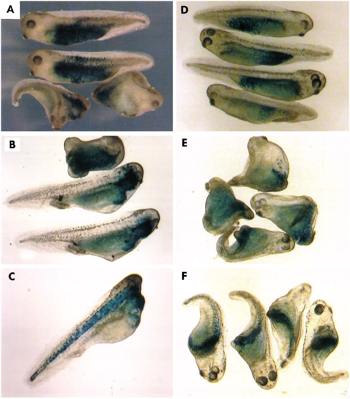
Fig. 6. Lineage tracing reveals the formation of dorsal and anterior structures by the progeny of a single blastomere injected with Xdsh mRNA.
Cleavage-stage (8-32 cells) embryos were coinjected with 0.4 ng of Xdsh mRNA and 0.2 ng of b-gal RNA or with b-gal RNA alone. After 2
days of development embryos were fixed, and stained for b-gal activity. (A) Normal embryos were injected at the 8- to 16-cell stage into a
ventrovegetal blastomere with b-gal RNA (two embryos on the top) or with b-gal and Xdsh mRNAs (bottom). (B,C) UV-treated embryos
rescued by Xdsh RNA and coinjected with b-gal RNA. (B) Staining is mainly in the pharyngeal endoderm and head mesenchyme of the fully
rescued embryos. A control embryo injected with b-gal RNA only is shown at the top. (C) In the partially rescued embryos (0.05-0.1 ng of
Xdsh RNA injected), the staining is in the notochord and pharyngeal endoderm. (D-F) Lineage tracing at the 32-cell stage. Embryos were
injected into D4 (tier 4) vegetal blastomere with b-gal RNA (D) or with b-gal and Xdsh mRNAs (E). (F) Xdsh and b-gal RNAs were
microinjected into C4 (tier 3) subequatorial blastomere. Staining is mainly in the notochord and anterior mesoderm. Image published in: Sokol SY et al. (1995) Copyright © 1995. Image reproduced with permission of the Publisher and the copyright holder. This is an Open Access article distributed under the terms of the Creative Commons Attribution License. Larger Image Printer Friendly View |