XB-IMG-135109
Xenbase Image ID: 135109
|
|||||||||||||||
|
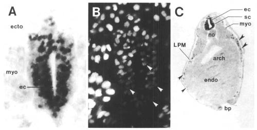
Fig. 5. Most neuronal precursor cells express XlHbox 6 in stage 24 spinal cord. Dorsal is uppermost in all panels.
(A) Section at high power magnification immunostained with anti-XlHbox 6 antibodies. (B) Same section counterstained
with Hoechst 33258 viewed under dark field illumination. Note that nearly all nuclei appear to be darkly stained in panel A
and that the Hoechst fluorescence in these nuclei is quenched by the immunostaining reaction product. (C) XlHbox 6
expression in lateral plate mesoderm. Transverse section of stage 25-26 tadpole decorated with XlHbox 6 antibodies and
overstained to show more clearly the less intense staining in the lateral plate mesoderm nuclei. Abbreviations:
ecto, ectoderm; ec, ependymal canal; sc, spinal cord; myo, myotome; LPM, lateral plate mesoderm; endo, endoderm;
arch, archenteron; no, notochord, remains of blastopore. Image published in: Wright CV et al. (1990) Copyright © 1990. Image reproduced with permission of the publisher and the copyright holder. This is an Open Access article distributed under the terms of the Creative Commons Attribution License.
Image source: Published Larger Image Printer Friendly View |