XB-IMG-135571
Xenbase Image ID: 135571
|
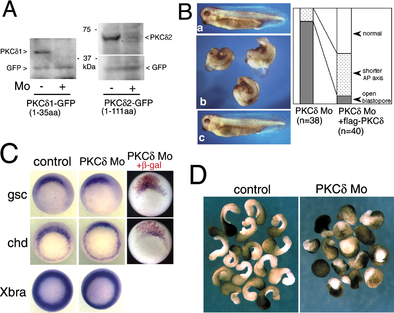
Figure 3.
PKCδ antisense morpholino also blocked gastrulation movements. (A) Morpholino oligonucleotides (MOs) for PKCδ1 and PKCδ2 inhibited the translation of mRNA that had the corresponding sequences. RNA encoding GFP-tagged PKCδ and unrelated GFP were coinjected with or without each MO. PKCδ1 (left panel) and PKCδ2 MO (right panel) blocked the production of each GFP-tagged PKCδ, but unrelated GFP was not affected. (B) Control MO or PKCδ MO (20 ng each) was injected into four-cell embryos (panels a,b, respectively). The PKCδ MO caused a gastrulation-defective phenotype that was indistinguishable from that of PKCδδC-injected embryos. This phenotype was rescued by 1 ng of full-length PKCδ1 RNA (panel c). (C) In situ hybridization of early gastrula embryos probed with chordin (chd), goosecoid (gsc), and Xbra. The left and middle panels show 20 ng of control or PKCδ MO was injected into all four blastomeres of four-cell embryos. (Right) To trace the cell lineage, mRNA encoding β-gal with a nuclear localization signal was coinjected with PKCδ MO into two dorsal blastomeres of four-cell embryos. (D) PKCδ MO inhibited the elongation of dorsal marginal zone explants. Twenty nanograms of MO were injected into the two dorsal blastomeres of four-cell embryos. Image published in: Kinoshita N et al. (2003) Copyright © 2003. Image reproduced on with permission of the Publisher, Cold Spring Harbor Laboratory Press. This is an Open Access article. Larger Image Printer Friendly View |