XB-IMG-135734
Xenbase Image ID: 135734
|
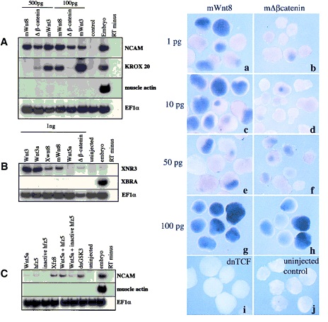
Figure 1. Wnt ligands, Frizzled receptors,
and signaling components induce
neural tissue in Xenopus ectodermal explants.
Either mWnt3, Xwnt3a, Xwnt8,
mWnt8, or mDb-catenin (500 pg or 100
pg) were injected into one-cell Xenopus
embryos. Ectoderm was removed at blastula
stage, and RNA extracted at late
neurula stage (20) (A,C) gastrula stage
(10.5) (B) or for analysis by RT-PCR. (A)
Injected ectoderm analyzed at neurula
stage for expression of the general neural
marker, NCAM, the hindbrain marker,
krox-20, the muscle-specific marker,
muscle actin, and the ubiquitously expressed
internal control, EF1a. (B) Injected
ectoderm analyzed at the gastrula
stage for the expression of the Wnt inducible
gene, Xnr3, the general mesodermal
marker, Xbra, and EF1a. (C) Ectoderm
injected with 1 ng each of the following:
Xwnt5a; human frizzled 5
(hfz5), a mutant human frizzled 5; Xenopus
frizzled 8 (Xfz8); dnGSK3; and combinations
of Xwnt5a, with either the
wild-type hfz5 or mutant hfz5. In A–C,
RNA from noninjected ectoderm, from
whole embryos treated with reverse
transcriptase, and from whole embryos
treated without reverse transcriptase
were used as controls. (D) Ectoderm injected
with 1, 10, 50, or 100 pg of either
mWnt8 (a,c,e,g) or mDb-catenin (b,d,f,h)
aged until stage 20 and analyzed by in
situ hybridization for Nrp1. Controls in
this experiment include ectoderm injected
with 1 ng of dnTCF (i) and uninjected
ectoderm (j). Image published in: Baker JC et al. (1999) Copyright © 1999. Image reproduced on with permission of the Publisher, Cold Spring Harbor Laboratory Press. This is an Open Access article. Larger Image Printer Friendly View |