XB-IMG-136012
Xenbase Image ID: 136012
|
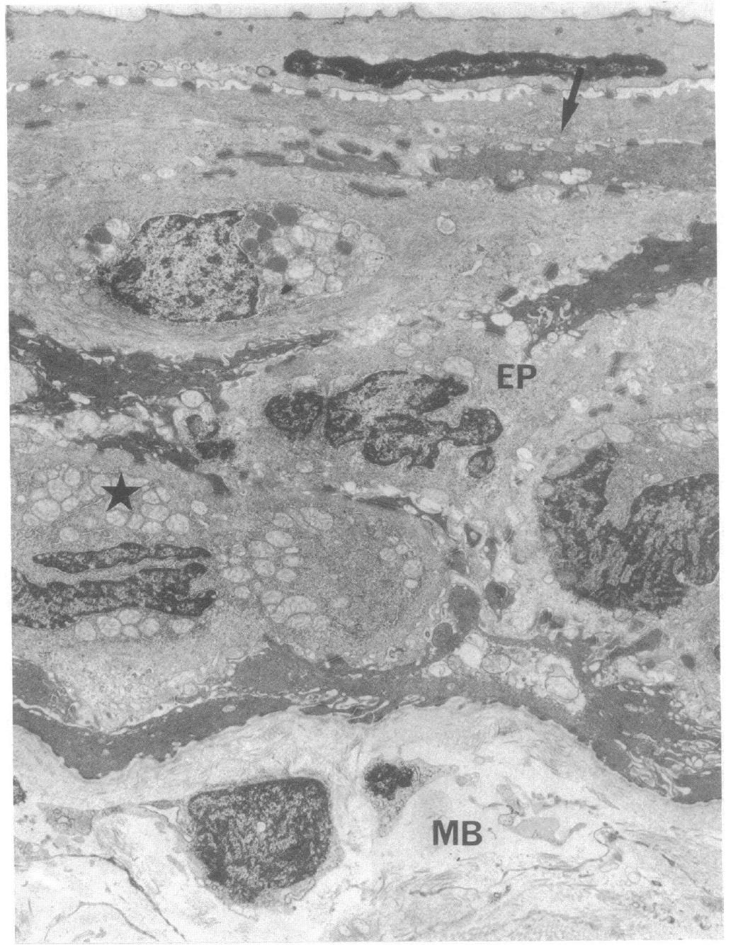
FIG. 4. Ultrastructure of the wound epithelium (EP). The cytoplasmic content is different in the outermost cells rich in cytokeratin
tonofilaments (arrow) as compared with the cells located below (star). There, in addition to intermediate filaments, mitochondria and free
ribosomes are quite prominent, suggesting high synthetic activity. Note the changes in size and electron density of the nucleus from the basal
region toward the surface of the epithelium where c-myc transcription is no longer observed (MB, mesenchymal cells). (x 7360). Image published in: Géraudie J et al. (1990) Copyright © 1990. Image reproduced with permission of the publisher and the copyright holder. This is an Open Access article distributed under the terms of the Creative Commons Attribution License. Larger Image Printer Friendly View |