XB-IMG-137169
Xenbase Image ID: 137169
|
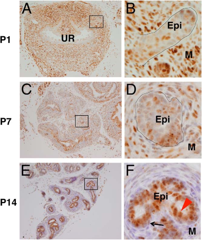
Figure 1. Bmp signaling was enhanced with the prostatic epithelial cell differentiation. A and B, Coronal sections of caudal body of ICR mice at P1; pSmad1/5/8 was weakly detected in the immature prostatic epithelia. C–F, Coronal sections of AP at P7 (C and D) and P14 (E and F). E and F, pSmad1/5/8 signal was more prominently observed in the differentiated luminal cells (F, red arrowhead) than that of the basal cells (F, black arrow) in the AP. UR, urethra. Epi, epithelia; M, mesenchyme. Scale bars, 100 μm. Image published in: Omori A et al. (2014) Copyright © 2014 by the Endocrine Society. This image is reproduced with permission of the journal and the copyright holder. This is an open-access article distributed under the terms of the Creative Commons Attribution license Larger Image Printer Friendly View |