XB-IMG-137207
Xenbase Image ID: 137207
|
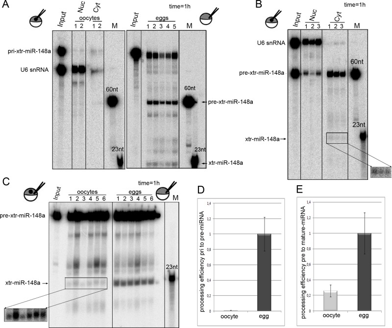
FIGURE 2:. Drosha and Dicer processing are enhanced upon maturation of oocytes to eggs. (A) pri-xtr-miR-148a is not processed when injected into oocyte nuclei (oocyte) but gets processed in eggs. RNA was injected in individual oocyte nuclei or eggs with U6 as a nuclear retention marker. RNAs were reextracted from corresponding single nuclei (Nuc) and cytoplasm (Cyt) of oocytes or from whole eggs. Minor processing of the miRNA could be detected in oocytes, whereas efficient processing to pre- and mature xtr-miR-148a was seen in eggs. (B) Nuclear export and processing of pre-xtr-miR148a in oocytes. Pre-xtr-miR-148a injected into oocyte nuclei is exported to the cytoplasm and processed to mature miR-148a. (C) Dicer processing is enhanced in eggs. Cytoplasmic injection of pre-xtr-miR-148a into oocytes shows moderate processing to mature miR-148a. The same amount of pre-xtr-miR148a injected into eggs is more efficiently processed. (D) Quantification of blot shown in A. The amount of processed RNA was quantified over the amount of input RNA. This shows a strong increase in Drosha cleavage in eggs. (E) Quantification of blot shown in C confirms a fivefold increase in Dicer processing. For A–C, incubation time was 1 h. Image published in: Muggenhumer D et al. (2014) © 2014 Muggenhumer, Vesely, et al. This image is reproduced with permission of the journal and the copyright holder. This is an open-access article distributed under the terms of the Creative Commons Attribution-NonCommercial-ShareAlike license Larger Image Printer Friendly View |