XB-IMG-137994
Xenbase Image ID: 137994
|
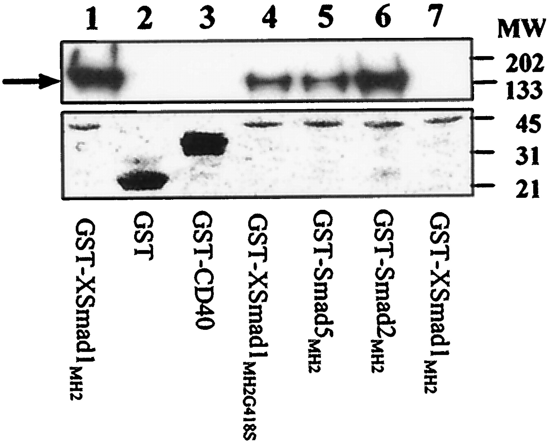
FIG. 4. In vitro association of SIP1 with the MH2 domains of
different Smad proteins. Myc-tagged full-size SIP1 protein was expressed
in COS1 cells. Upper panel, equal amounts of the same cell
extract were used in lanes 1–6. The 145-kDa SIP1 protein (indicated by
the arrow) was efficiently pulled down from this cell lysate using the
different GST-Smad fusion proteins (lanes 1 and 4–6; visualization is
by Western blotting using anti-Myc antibody) but not by an unrelated
GST-fusion protein (GST-CD40; lane 3) and GST (lane 2). GST fusions
included the MH2 domains of XSmad1(G418S) (lane 4), mouse Smad5
(lane 5), mouse Smad2 (lane 6), and wild type XSmad1 (lane 1), respectively.
Lane 7 provides a negative control with proteins pulled-down by
GST-XSmad1 from a cell lysate of mock-transfected cells. Lower panel,
estimate of the amount of GST-fusion proteins used in the pull-down
experiments by Ponceau S staining of the used blot. Lower amounts of
GST-Smad fusion proteins were used in lanes 1 and 4–7. Image published in: Verschueren K et al. (1999) Copyright © 1999. Image reproduced with permission of the publisher and the copyright holder. This is an Open Access article distributed under the terms of the Creative Commons Attribution License. Larger Image Printer Friendly View |