XB-IMG-138149
Xenbase Image ID: 138149
|
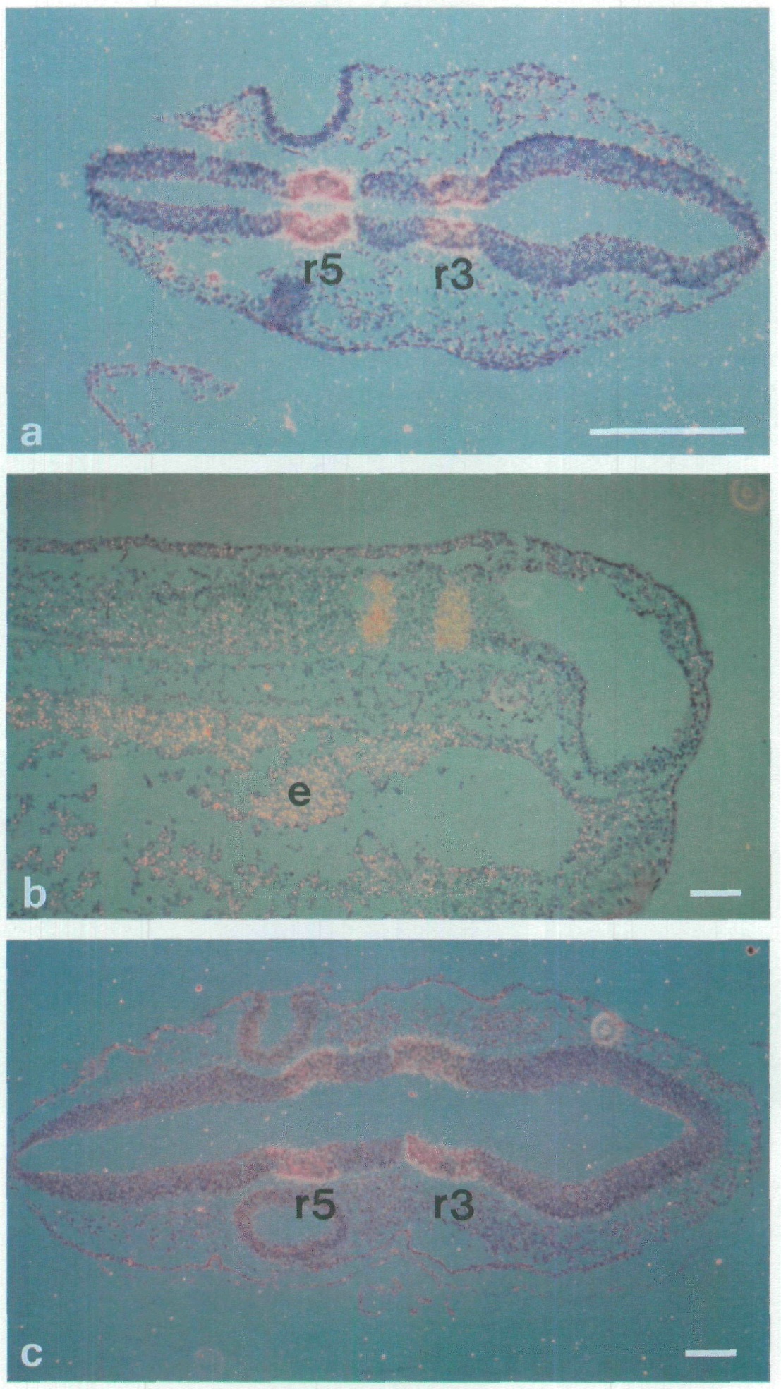
Fig. 2. Conserved patterns of
Krox-20 expression in mouse,
Xenopus and chick. In situ
hybridisation was carried out
using appropriate
homologous Krox-20 probes
as described (Wilkinson and
Green, 1990). (a) 9.5 day
mouse embryo; (b) stage 28
Xenopus embryo; (c) stage 15
chick embryo, r,
rhombomere. The apparent
signal in the endoderm (e) of
the Xenopus embryo is due
to the refraction of light by
yolky cells, not the
hybridisation of probe.
Anterior is to the right in all
photographs. Bar=100/«m. Image published in: Nieto MA et al. (1991) Copyright © 1991. Image reproduced with permission of the publisher and the copyright holder. This is an Open Access article distributed under the terms of the Creative Commons Attribution License.
Image source: Published Larger Image Printer Friendly View |