XB-IMG-138216
Xenbase Image ID: 138216
|
|
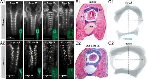
Figure 2. Structural deformations of individual vertebrae after UL. A, Top view of calcein-stained cartilaginous/bony elements of the vertebral column at subsequent stages of premetamorphic and postmetamorphic development in controls (A1) and after UL (A2, Post-UL) at different postlesional intervals (15â47 d); insets show the respective developmental stages; yellow arrowheads and red asterisks indicate deformations of the vertebral column and side of the UL, respectively. B, Cross-section (10 μm) through the rostral tail region at the level of myotome 4 in a stage 57 control tadpole (B1) and 20 d post-UL (B2); specific colorimetric staining of cartilage with Alcian blue and other tissue with hematoxilin-eosin differentiated vertebral elements such as cartilage (car), muscle (mus), spinal cord (sc), and the chorda dorsalis (cho). C, Stacked schematic outlines of cross-sectioned cartilaginous vertebral tissue at the level of myotome 4 (blue lines) of a control (C1) and 20 d post-UL (C2); data are from the material shown in B1 and B2, respectively, and represent a superimposed overlay of 10 successive 10 μm thick sections centered on the spinal cord. Image published in: Lambert FM et al. (2013) Copyright © 2013. OA ARTICLE, images redisplayed under a Creative Commons license. Larger Image Printer Friendly View |