XB-IMG-138217
Xenbase Image ID: 138217
|
Figure 3.
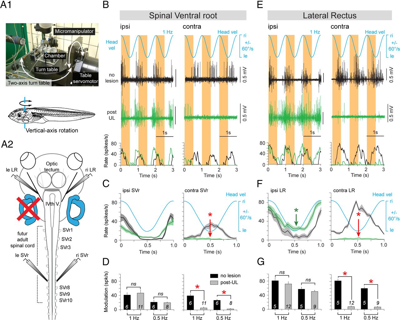
Postlesional changes of SVr and extraocular motor activity during vertical-axis vestibular stimulation in stage 55â57 Xenopus tadpoles. A, Computer-controlled two-axis turntable (A1) allowed sinusoidal rotations around vertical (A1, bottom) and horizontal axis of semi-intact preparations from control animals and after a UL on the left side (A2). BâG, Spike discharge and firing rate modulation of an SVr (B) and LR (E) on the ipsilesional side (left columns) and contralesional side (right columns) during sinusoidal head rotation at 1 Hz (±60°/s; blue traces; Headvel) in a control (no lesion, black traces) and 6 weeks post-UL (green traces). Mean discharge rate over one cycle (n = 30; ±SE; shaded area in each plot) of the ipsilesional and contralesional SVr (C) and LR (F) in a control (black curves) and after UL (green curves) with respect to Headvel. Average modulation (±SE) of ipsilesional and contralesional SVr (D) and LR (G) in response to 1 and 0.5 Hz vertical-axis sinusoidal head rotations, respectively. The significance of difference in the peak firing rate of the respective nerve roots between controls and the postlesional group was tested with the MannâWhitney U test for unpaired parameters (*p ⤠0.05; n.s.). The number of animals are indicated; ipsi, ipsilesional side; contra, contralesional side; ri, right, le, left; IVth V, IVth ventricle. Image published in: Lambert FM et al. (2013) Copyright © 2013. OA ARTICLE, images redisplayed under a Creative Commons license. Larger Image Printer Friendly View |