XB-IMG-138218
Xenbase Image ID: 138218
|
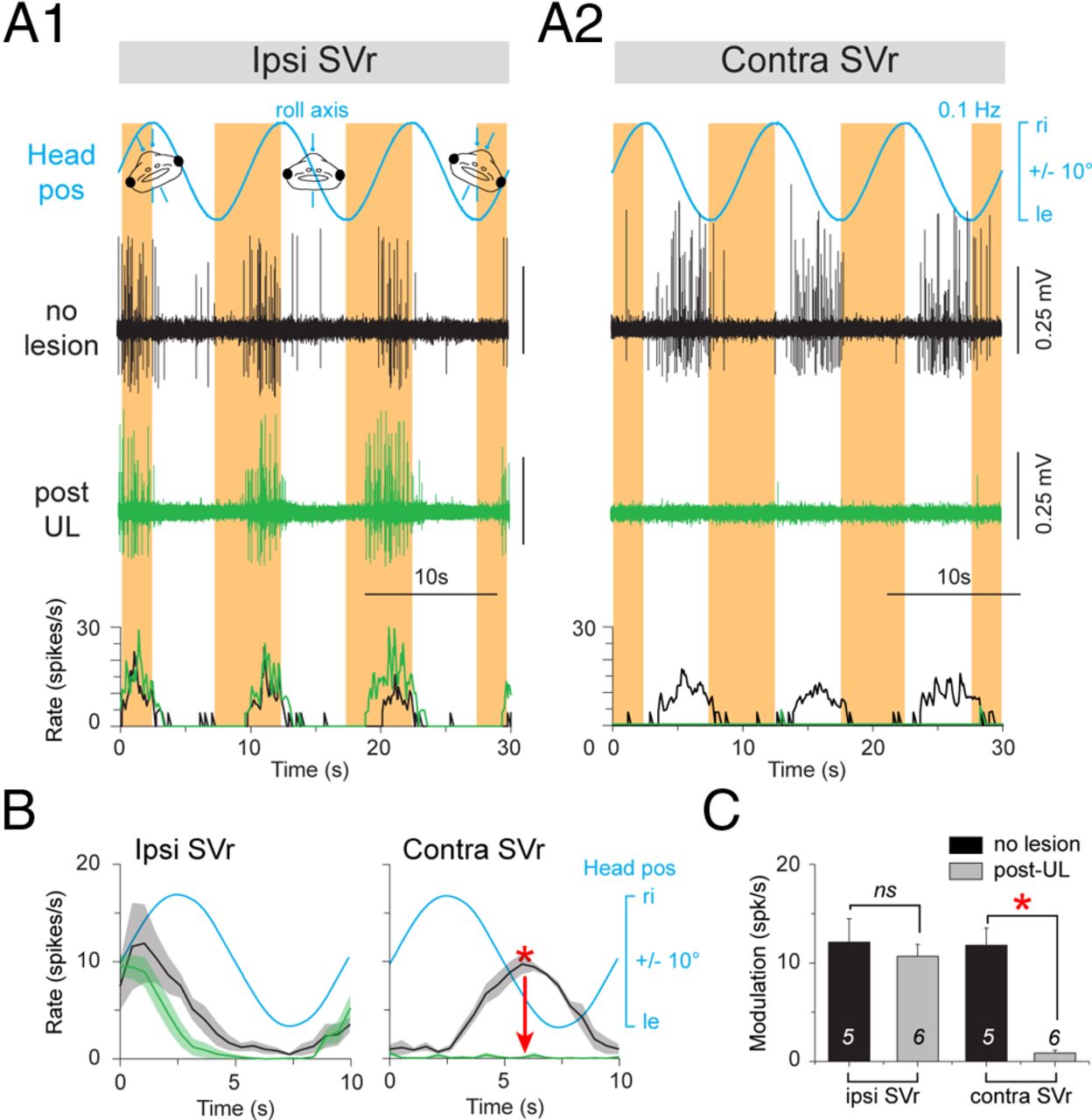
Figure 4. Postlesional changes of SVr activity during horizontal-axis vestibular stimulation in a stage 57 larval Xenopus. A, Spike discharge and firing rate modulation of the SVr on the ipsilateral side (A1) and contralesional side (A2) during sinusoidal left-right horizontal-axis head rotation (roll-axis) at 0.1 Hz (±6°/s; blue traces, Headpos) in a control (no lesion, black traces) and 6 weeks post-UL (green traces). B, Mean discharge rate over one cycle (n = 30; ±SE; shaded area in each plot) of the ipsilesional and contralesional SVr in a control (black traces) and after UL (green traces) with respect to Headpos. C, Average modulation (±SE) of ipsilesional and contralesional SVr during 0.1 Hz roll-axis sinusoidal rotations before (n = 5) and after (n = 6) UL. The significance of difference in the peak firing rate of the respective ispilesional and contralesional SVr between controls and the postlesional group was tested with the Mann–Whitney U test for unpaired parameters (*p ≤ 0.05; n.s.). ipsi indicates ipsilesional side; contra, contralesional side; ri, right, le, left; IVth V, IVth ventricle. Image published in: Lambert FM et al. (2013) Copyright © 2013. OA ARTICLE, images redisplayed under a Creative Commons license. Larger Image Printer Friendly View |