XB-IMG-138291
Xenbase Image ID: 138291
|
||||||||||
|
AFigure
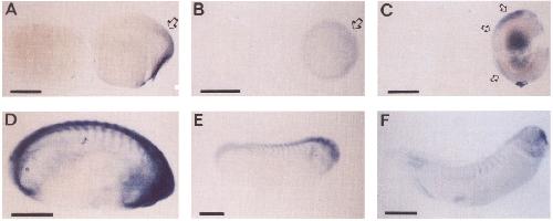
4. Kv2.2 transcripts are present in excitable tissues of developing Xenopus embryos. A-C, In each view, the embryo on the left was hybridized to
a digoxigenin sense control cRNA probe, whereas the embryo on the right was hybridized to a Kv2.2 antisense cRNA probe (scale bars: A, C, 0.5 mm;
B, 1 mm).A, Early gastrula stage embryo (stage 9; 7 hr after fertilization). Staining is apparent in the dorsal lip of the blastopore (arrow). B, Late gastrula
stage (stage 12; 14 hr). Kv2.2 staining localizes to dorsal ectoderm and presumptive neural tissue (arrow). C, Neurula stage (stage 19; 20 hr). Kv2.2 mRNA
is localized along the entire neural tube (arrows). Rostra1 is up; dorsal is to the leff. The dark shadow in the gut of the embryo is attributable to incomplete
clearing of the embryo. D-F, In these lateral views, rostra1 is to the right, and dorsal is up (scale bars: D-F, 0.5 mm). Sense controls for these older stages
are not shown but are similar to those shown in A-C. D, Expression of Kv2.2 mRNA in the early tail bud embryo (stage 23; 1 d). Staining is present in
the brain, spinal cord, and anterior midsomite regions. E, A similar pattern of Kv2.2 mRNA expression is observed 6 hr later in the stage 26 embryo, except
that the signal in the midsomite regions has extended caudally. F, In the stage 35 embryo (2 d), Kv2.2 staining is faint in the majority of the brain and
spinal cord but still visible in the midsomite regions. The dark signal at the tip of the embryo corresponds to the forebrain. Image published in: Burger C and Ribera AB (1996) Copyright © 1996. This image is reproduced with permission of the publisher and the copyright holder. This is an Open Access article distributed under the terms of the Creative Commons Attribution License.
Image source: Published Larger Image Printer Friendly View |