XB-IMG-138475
Xenbase Image ID: 138475
|
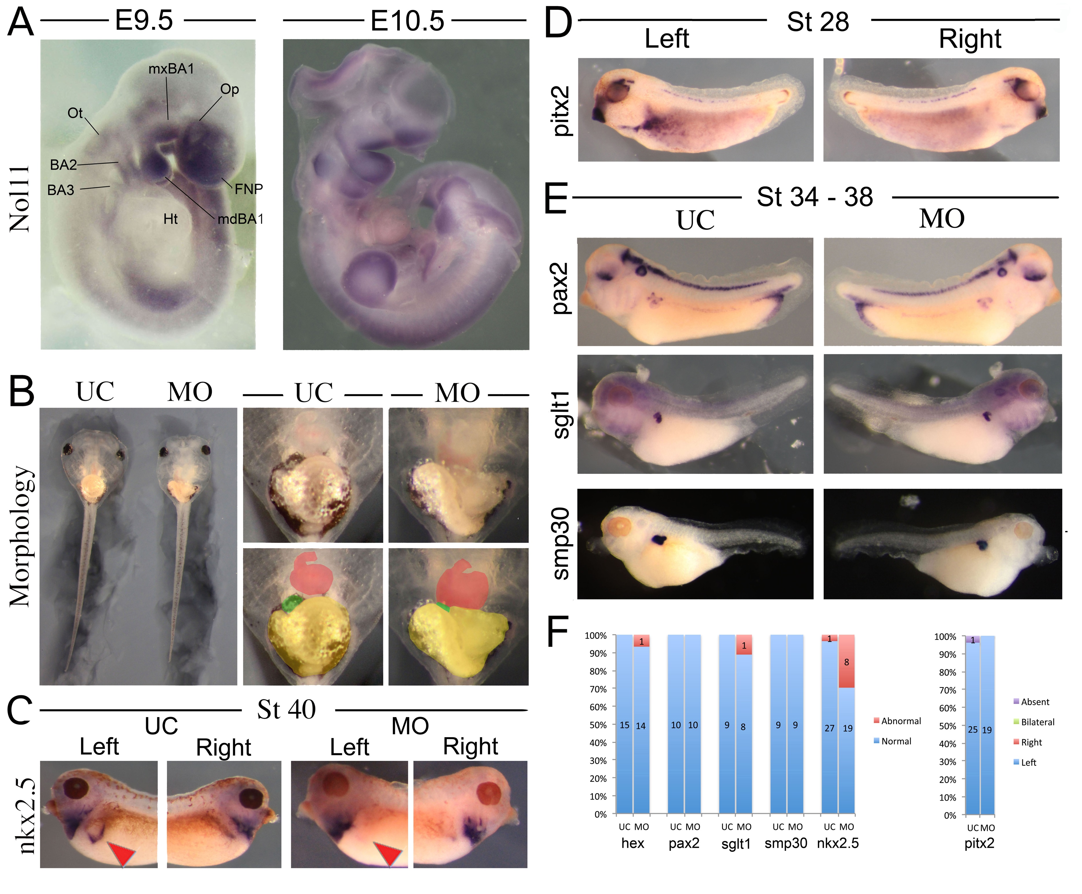
S1 Fig. nol11 in vertebrate development.
A) Whole mount in situ hybridization of digoxigenin labelled Nol11 probe in E9.5 and E10.5 mouse embryos (E10.5 sample shown has been hemisected). BA, branchial arch; FNP, frontonasal prominence; Ht, heart; mdBA1, mandibular BA1; mxBA1, maxillary BA1; Op, optic placode; Ot, otic placode. B) Gross morphology of stage 45 nol11 morphants. Gut morphology is abnormal in morphants, while organ situs appears largely normal relative to wild type controls. Heart (red), gall bladder (green) and gut (yellow) are pseudocoloured in lower right panels. C) Left sided expression nkx2.5 is reduced or absent in the splenic anlage of a subset of nol11 morphants (compare red arrowheads). D) Example of the normal sided pitx2 expression present in nol11 knocked down embryos. E) Kidney development appears largely intact in nol11 MO treated side compared to control side. F) Quantification of number of embryos displaying the described phenotypes.
doi:10.1371/journal.pgen.1005018.s001 Image published in: Griffin JN et al. (2015) Image reproduced on Xenbase with permission of the publisher and the copyright holder. This image is reproduced with permission of the journal and the copyright holder. This is an open-access article distributed under the terms of the Creative Commons Attribution license
Image source: Published Larger Image Printer Friendly View |