XB-IMG-139032
Xenbase Image ID: 139032
|
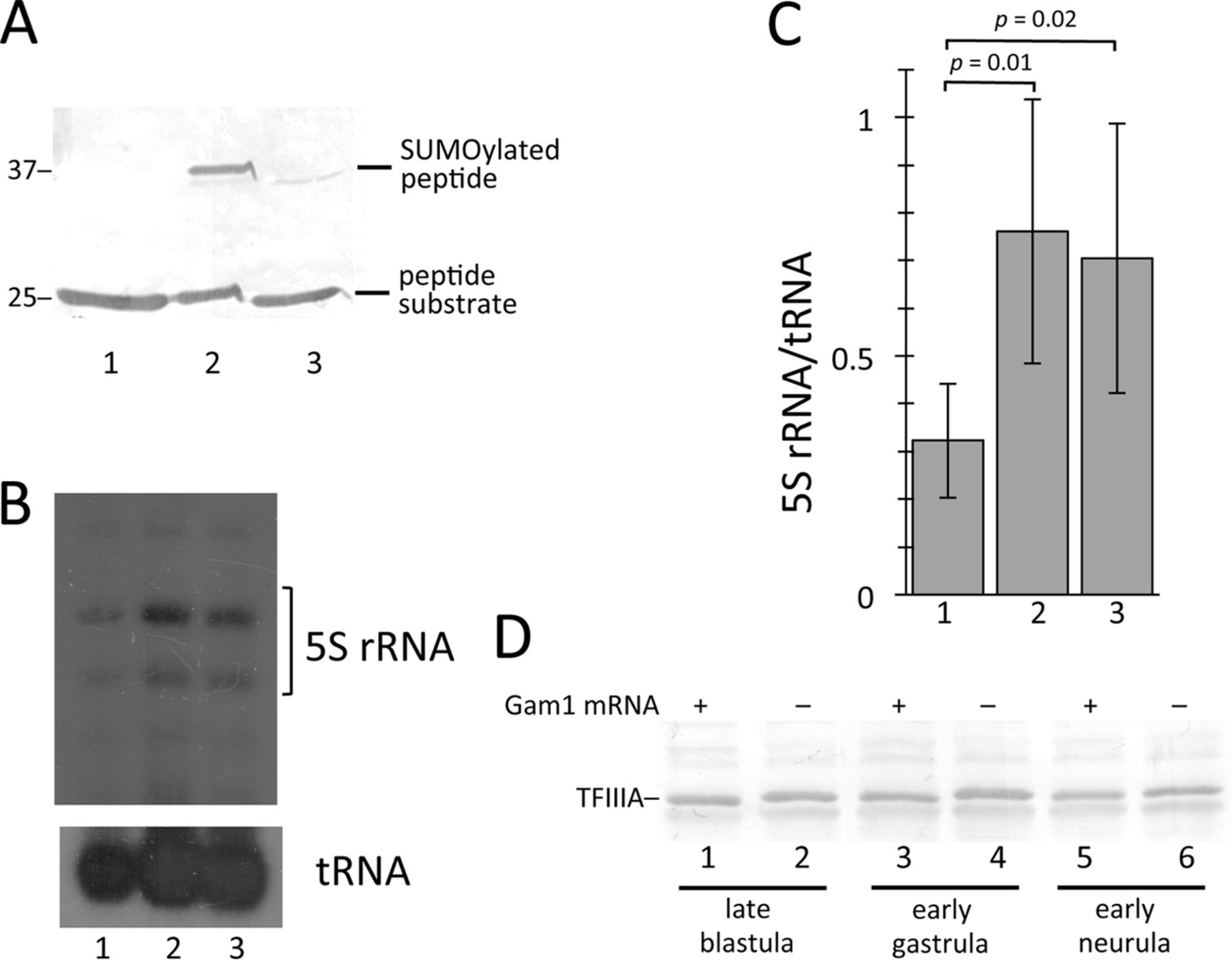
FIGURE 4.
Depletion of SUMOylation activity prevents repression of oocyte 5 S rRNA genes. A, SUMO activation (E1) activity in extract prepared from Gam1-injected embryos was measured by Western blot analysis using a 25-kDa SUMO substrate peptide. Lane 1, peptide alone; lane 2, peptide incubated with extract from water-injected (control) embryos; lane 3, peptide incubated with extract from Gam1-injected embryos. The reactions in lanes 2 and 3 also contained E2 (Ubc9) enzyme, SUMO1, and ATP. Positions of molecular weight markers (in kilodaltons) are indicated. B, one-cell embryos were injected with [32P]UTP and TFIIIA mRNA, which activates transcription of the oocyte-type genes after the midblastula transition, or Gam1 mRNA. RNA was isolated at early gastrula stage, and three embryo equivalents were analyzed by electrophoresis/autoradiography. Lane 1, control embryos injected with [32P]UTP only; lane 2, injection with TFIIIA mRNA (7 ng); lane 3, injection with Gam1 mRNA (5 ng). C, autoradiographs for four experiments were scanned, and 5 S rRNA was quantitated relative to tRNA using ImageJ software. The ratio is given in arbitrary units. Error bars indicate mean ± S.D. D, one-cell embryos were injected with Gam1 mRNA (lanes 1, 3, and 5) or water (lanes 2, 4, and 6). Whole cell extract was prepared, and four embryo equivalents were analyzed by Western blot developed with TFIIIA antibody. Image published in: Malik MQ et al. (2014) Copyright © 2014. Image reproduced with permission of the publisher and the copyright holder. This is an Open Access article distributed under the terms of the Creative Commons Attribution License. Larger Image Printer Friendly View |