XB-IMG-140013
Xenbase Image ID: 140013
|
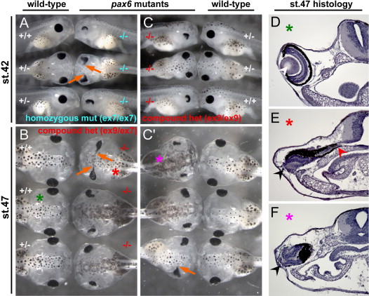
Fig. 5. Phenotypes of Xenopus pax6 mutants at tadpole stages. (A) Phenotypes of F2 embryos at stage 42 from an intercross of F1 heterozygotes carrying a 13-bp deletion in exon 7. −/−, homozygous mutant; +/−, heterozygous; +/+, wild-type. These are siblings of embryos shown in Fig. 4B. (B) Phenotypes of F2 embryos at stage 47 from cross between an F1 female carrying a 1-bp deletion ( #2) in exon 9 and an F1 male carrying a 13-bp deletion in exon 7. −/−, compound heterozygous mutant (genotype is ex9 δ1-bp#2/ex7 δ13-bp); +/−, heterozygous (genotype, +/ex7 δ13-bp); +/+, wild-type. These are siblings of embryos shown in Fig. 4D. (C and C′) Phenotype of F2 embryos at stage 42 (C) and stage 47 (C′) from mating of an F1 female carrying a 1-bp deletion (#2) in exon 9 and F1 male carrying a 1-bp deletion (#3) in exon 9. Embryos shown in C (mixture of lateral and dorsal views) are the same embryos as shown in C′ (all dorsal views) at the same axial position, the only difference being their ages. −/−, compound heterozygous mutant (genotype is ex9 δ1-bp#2/ex9 δ1-bp#3); +/−, heterozygous wild-type (genotype, ex9 δ1-bp#2/+ for top embryo, +/ex9 δ1-bp#3 for second embryo); +/+, homozygous wild-type. These are siblings of embryos shown in Fig. 4C. Orange arrows show the eye fused to the brain, which is often, but not always, seen in pax6 mutants regardless of genotype. (D–F) Cross sections of the eye region of embryos shown in (B) and (C′). Corresponding embryo and section are marked by the same colored asterisks. Wild-type embryos (D) have well organized retina and lens (white arrowhead), whereas mutants (E and F) do not have lens and the retina is disorganized (black arrowheads). By this stage, mutant eyes were separated from the brain in some embryos (F) or still connected to the brain in other embryos (E, red arrowhead). (For interpretation of the references to color in this figure legend, the reader is referred to the web version of this article.) Image published in: Nakayama T et al. (2015) Copyright © 2015. Image reproduced with permission of the Publisher, Elsevier B. V. Larger Image Printer Friendly View |