XB-IMG-140018
Xenbase Image ID: 140018
|
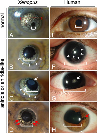
Fig. 10. Comparison of aniridia-like frog eyes with human aniridic eyes. For purposes of comparison the images of froglet eyes shown here are approximately half the size of the human eyes. Actual froglet corneal diameter is approximately one-tenth that of human. Normal frog (A) and human (E) eyes have small pupil (indicated by white brackets) and relatively large, pigmented iris. Aniridia-like mutant froglets (BâD) and human aniridia patients (FâH) show variable eye phenotypes including reduced (BâD, H) or absent (F and G) iris tissue. Remaining iris tissue may show focal losses (red arrows D and H). The lenses may be clear (B and F) or have a focal cataract (white arrows in C and G). Also, both frog and human eyes may show peripheral clouding of the cornea due to keratopathy (white arrowheads in B, C and F). (For interpretation of the references to color in this figure legend, the reader is referred to the web version of this article.) Image published in: Nakayama T et al. (2015) Copyright © 2015. Image reproduced with permission of the Publisher, Elsevier B. V. Larger Image Printer Friendly View |