XB-IMG-140021
Xenbase Image ID: 140021
|
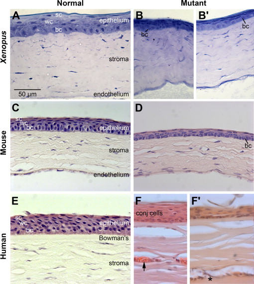
Fig. 13. Xenopus pax6 mutant exhibits corneal defects similar to those observed in Pax6 mutant mice and human aniridia. Histology of the cornea of wild-type (Normal) and Pax6 mutant (Mutant) frog (Xenopus), mice (Mouse), and human (Human). (A) Wild-type froglet. (B) pax6 Hypomorphic froglet. (C) 8-Week-old adult wild-type mouse (D) 8-Week-old adult Pax6Sey-Neu/+ mouse. (E) 70-Year-old human. The cornea did not show clinical signs of keratopathy. (FâFâ²) 49-Year-old individual with classical aniridia. The patient was diagnosed with stage III keratopathy and exhibited full conjunctivalization of the cornea. Arrow denotes blood vessel. Asterisk denotes area of cellular infiltration in the superficial corneal stroma. The samples here were of peripheral cornea. Note: the endothelial layer was not captured in these images of human cornea. bc, basal cells; conj, conjunctival cells, sc, superficial cells; wc, wing cells. Scale bar in panel A applies to panels (BâF). (For interpretation of the references to color in this figure legend, the reader is referred to the web version of this article.) Image published in: Nakayama T et al. (2015) Copyright © 2015. Image reproduced with permission of the Publisher, Elsevier B. V. Larger Image Printer Friendly View |