XB-IMG-140030
Xenbase Image ID: 140030
|
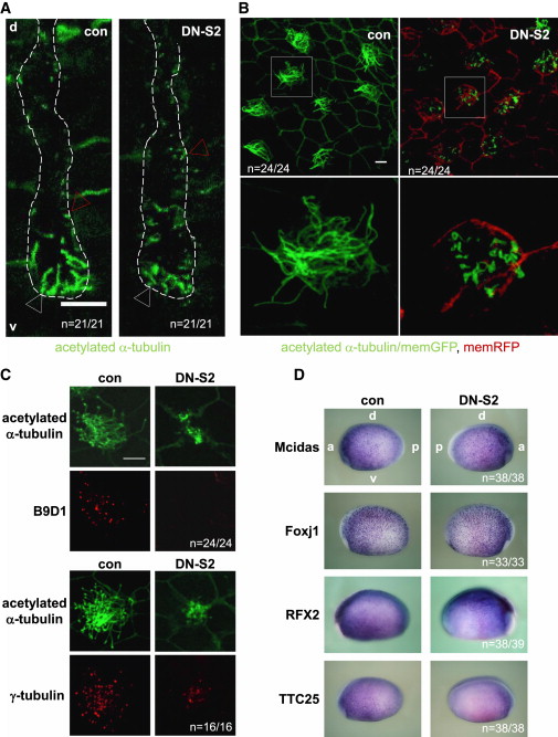
Figure 4. TGF-b Signaling Controls the Length of Motile Neural and Epidermal Cilia and Affects the Transition Zone in a Mcidas/Foxj1/RFX2-Independent Manner. (A) Shortened FL cilia in the neural tube. DN-S2 RNA (4 ng) was injected into the dorsal animal region of four-cell embryos, which were fixed at stage 26. Following staining, 100 mm transverse
sections of specimens were prepared. Red and white arrowheads indicate examples of non-motile monocilia and FL-cilia, respectively. White dots
indicate the lumen of the neural tube. The scale bar represents 10 mm. d, dorsal side, v, ventral side. (B) Shortened cilia on epidermal MCC. DN-S2 RNA (4 ng) with memRFP RNA or memGFP RNA was injected into the left side or the right side, respectively. Embryos were fixed at stage 26.
Images from the left (DN-S2) or right (con) lateral side were taken. Pictures in the lower row were magnified from the white square indicated in each
sample. The scale bar represents 10 mm. (C) g-tubulin, but not B9D1, was detected in DN-S2-injected MCC cilia. DN-S2 RNA (4 ng) with memGFP RNA or memGFP RNA alone was injected into the left or right side, respectively. Images from the left (DN-S2) or right (con) lateral side were taken. Scale bar represents 10 mm. (D) Mcidas, Foxj1, RFX2, and TTC25 expression in DN-S2-injected embryos at stage 17 by wholemount
in situ hybridization. Images from the left (con) or right (DN-S2) lateral side were taken. Red-gal signals indicate the DN-S2-injected side.
a, anterior; p, posterior; d, dorsal; v, ventral. âânââ indicates number of embryos. Statistical analyses were carried out using Excel using SEM and Studentâs t test to calculate p values (*p % 0.05, **p % 0.005, ***p % 0.0005, ****p % 0.00005; n.s., not significant). Image published in: Tözser J et al. (2015) Copyright © 2015. Image reproduced with permission of the Publisher and the copyright holder. This is an Open Access article distributed under the terms of the Creative Commons Attribution License.
Image source: Published Larger Image Printer Friendly View |