XB-IMG-144948
Xenbase Image ID: 144948
|
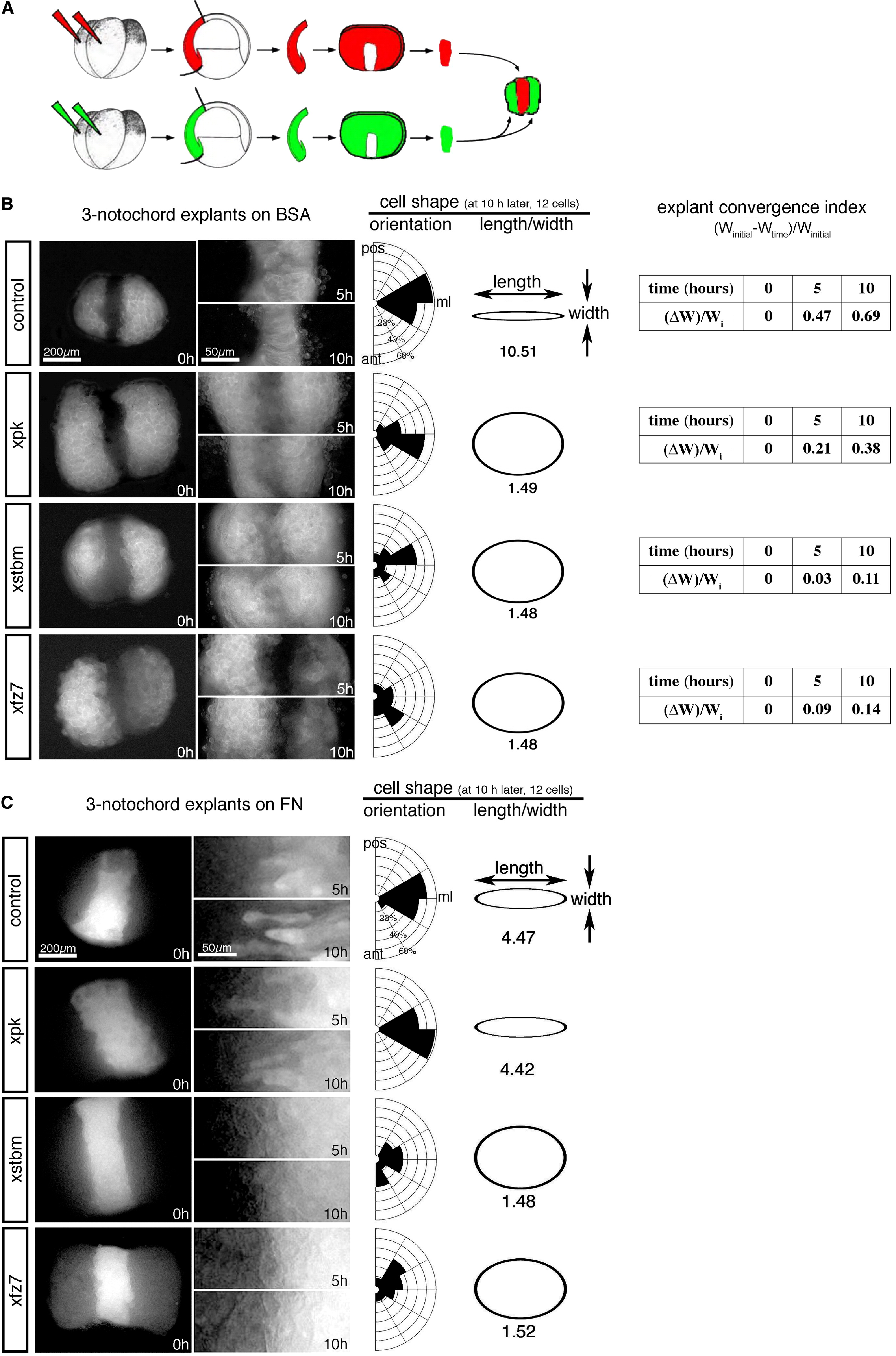
Figure 2.
Cell Intercalation in Three-Notochord Explants
(A) Procedure for making three-notochord explants. Xstbm, Xpk, or Xfz7 mRNA with green tracer (GFP or gap43-GFP) or red tracer (Alexa594 dextran) was coinjected into two dorsal blastomeres of the 4-cell-stage embryo. Notochord sectors (without epithelium) were dissected from injected embryos at stage 10.
(B and C) Three-notochord sectors from different embryos were grafted together and cultured on either the BSA (B)- or fibronectin (C)-coated glass, exposing the deep cells to two fluorescent color time-lapse imaging. Control explants were made from embryos injected with GFP or gap43-GFP and Alexa594 dextran. The time elapsed is indicated at the bottom right. (B) On BSA, cells in control explants adopt bipolar shapes and intercalate deeply into the neighbor notochord to make a single big notochord. Xpk-overexpressing explants converged weakly. Xstbm- and Xfz7-overexpressed cells remain round and the explants never converge. (C) On fibronectin, cells in the central notochord of control explants adopt bipolar shapes and intercalate deeply. Likewise, cells within Xpk-overexpressing explants become bipolar and intercalate. In contrast, Xstbm- and Xfz7-overexpressed cells remain round and do not intercalate as in (B). ml, mediolateral.
Image published in: Goto T et al. (2005) Copyright © 2005. Image reproduced with permission of the Publisher, Elsevier B. V. Larger Image Printer Friendly View |