XB-IMG-145018
Xenbase Image ID: 145018
|
|
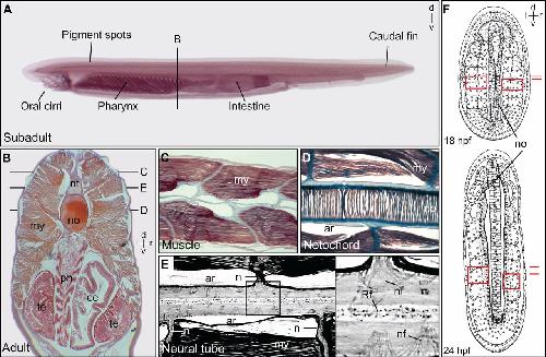
Fig. 5. Asymmetry in amphioxus. (A) Subadult animal (before differentiation of the gonads). Histological transverse (B) and longitudinal (C-E) sections of adult animals. The longitudinal sections were taken at the level of the dorsal muscle, dorsal to the neural tube (nt; C), at the level of the notochord (no; D) and at the level of the neural tube (E). Note the asymmetric body plan as reflected in the placement of the pharynx (ph), cecum (ce) and testes (te), and the alignment of muscles [myomeres (my)], nerve (n) and nerve fibers (nf). ar, fixation artifact; Rf, Reissner's fiber. (F) Reproduction of original drawings from Conklin's 1932 description of embryogenesis in amphioxus [reproduced with permission (Conklin, 1932)]. At 18 h post-fertilization (18 hpf, top), the somites are symmetrically aligned. However, by 24 hpf (bottom), somitogenesis has become out of register, as is obvious from the seventh somite onwards. The fourth (top) and seventh (bottom) somites are boxed in red. d, dorsal; l, left; r, right; v, ventral. Image published in: Blum M et al. (2014) Copyright © 2014. Image reproduced with permission of the publisher and the copyright holder. This is an Open Access article distributed under the terms of the Creative Commons Attribution License. Larger Image Printer Friendly View |