XB-IMG-145530
Xenbase Image ID: 145530
|
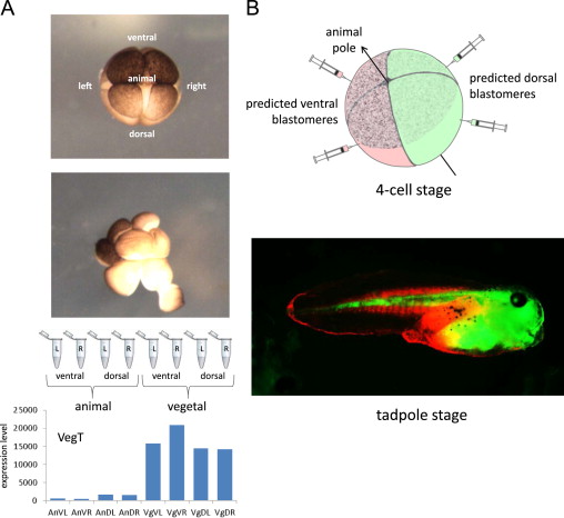
Fig. 2. A. Disassembling and sequencing the 8-cell stage embryo. Photograph of the intact 8-cell embryo, and during microsurgical disassembly; note lighter dorsal/animal blastomeres. Sequencing of single blastomeres from a single embryo, showing layout of displayed data for known vegetally enriched gene, VegT. B. Dye tracing experiment to confirm correct identification of dorsal–ventral axis at the 4-cell, and by extension at the 8-cell, stage. The dorsal/animal quadrant is identified by more lightly pigmented blastomeres. Image published in: De Domenico E et al. (2015) © 2015 The Authors. This image is reproduced with permission of the journal and the copyright holder. This is an open-access article distributed under the terms of the Creative Commons Attribution license Larger Image Printer Friendly View |