XB-IMG-147780
Xenbase Image ID: 147780

|
|
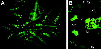
Figure 3. Pax-6/green fluorescent protein (GFP) transgenics show complex patterns of gene expression in later development, demarcating numerous subdomains within the brain, spinal cord, and sensory tissues. A: A group of Pax-6/GFP transgenic F2 siblings at late swimming tadpole stages (Nieuwkoop and Faber stage 43â45; Nieuwkoop and Faber, 1956). Note the consistency of GFP pattern and intensity between tadpoles. B: A high-magnification view of the head of a Pax-6/GFP primary transgenic tadpole at stage 46. Pax-6/GFP is expressed in the brain (br), eye (ey; GFP fluorescence is blocked in this image by the pigmented retina), and olfactory placodes (olf). Arrows point to the olfactory nerve (between olfactory placodes and brain) and optic nerve (between eye and brain), which show low, but detectable, levels of GFP fluorescence. Small clusters of cells on the skin, which may be part of the lateral line (Winklbauer, 1989), also express GFP. Image published in: Hirsch N et al. (2002) Copyright © 2002. Image reproduced with permission of the Publisher, John Wiley & Sons. Larger Image Printer Friendly View |
