XB-IMG-147888
Xenbase Image ID: 147888
|
|
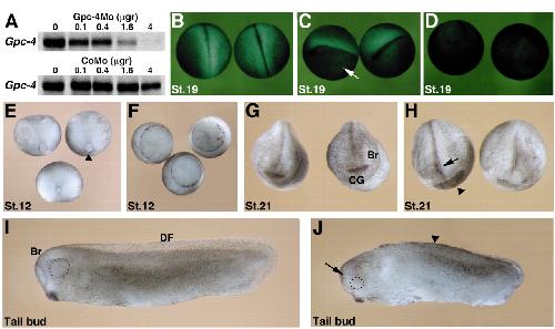
Fig 2. GPC4 is required for early embryonic development. (A) The Gpc4Mo inhibits translation of Gpc4 mRNA in vitro. Capped mRNA was in vitro translated in the presence of increasing amounts (indicated in μg) of Gpc4Mo (top panel) or CoMo (bottom panel). (B-D) Gpc4Mo specifically inhibits translation of Gpc4 transcripts in vivo. (B) Embryos injected with chimeric Gpc4GFP transcripts and CoMo. (C,D) Embryos injected with Gpc4GFP mRNA and Gpc4Mo in one blastomere (arrow in C) or both blastomeres (D). Injection of Gpc4Mo inhibits Gpc4GFP mRNA translation as evidenced by lack of GFP activity. (E-J) Two-cell embryos were injected with CoMo (E,G,I) or Gpc4Mo (F,H,J) and analysed at different developmental stages. (E,F) GPC4 functions during gastrulation. Dorso-vegetal view of stage 12 embryos. (E) Blastopore has closed in embryos injected with CoMo (stage 12). (F) Blastopore remains open in embryos injected with Gpc4Mo (stage 12). (G,H) GPC4 is required for anterior CNS development. Frontal view of stage 21 embryos. Embryos injected with Gpc4Mo (H) retain an open anterior neural tube (arrow) but develop a cement gland (arrowhead). (I,J) Side view of tailbud stage embryos. In contrast to control embryos (I), embryos injected with Gpc4Mo (J) are shorter, lack the dorsal fin and have small heads. Arrowhead in J points to the missing dorsal fin; the arrow indicates microcephaly. The developing eyes are encircled. CG, cement gland; Br, brain; DF, dorsal fin. Image published in: Galli A et al. (2003) Copyright © 2003. Image reproduced with permission of the publisher and the copyright holder. This is an Open Access article distributed under the terms of the Creative Commons Attribution License. Larger Image Printer Friendly View |