XB-IMG-149111
Xenbase Image ID: 149111
|
|||||||||||||||
|
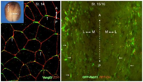
Fig. 2. Anteroposterior and mediolateral PCP in the Xenopus neural plate.En face views of the Xenopus neural plate shown for stage 14–16 embryos. Left, immunostaining for Vangl2 and ZO1 reveals anterior accumulation of Vangl2 in neural midline cells (arrows). Inset shows whole embryo view with indicated anteroposterior (A-P) and mediolateral (M-L) axes. Right, the middle area of the neural plate exhibits mediolateral planar polarity of the cells mosaically expressing GFP-Rab11 (green) and RFP-Diversin (red puncta) that are enriched at medial domains of cells in the lateral neural plate. Arrows mark cell polarization towards the midline (dotted line). Image published in: Sokol SY (2015) Copyright © 2015. This image is reproduced with permission of the publisher and the copyright holder. This is an Open Access article distributed under the terms of the Creative Commons Attribution License.
Image source: Published Larger Image Printer Friendly View |