XB-IMG-149335
Xenbase Image ID: 149335

|
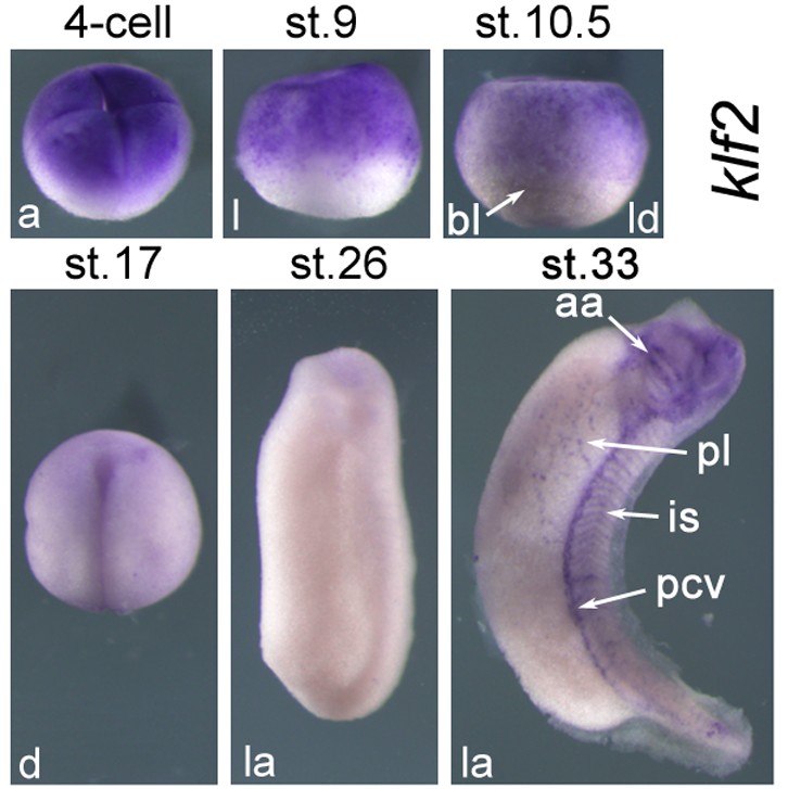
Fig. 2. A: Spatial expression patterns klf2, during Xenopus embryogenesis detected with whole mount in situ hybridization. a, animal view; aa, aortic arches; ad, adenohypophysial placode; an, anterior view, dorsal is up; ba, branchial arches; bl, blastopore lip; cg, cement gland; d, dorsal view, the anterior is up; da, dorsal anterior view; dv, dorsal vegetal view; ey, eye; fb, forebrain; fep, facial epibranchial placode; hb, hindbrain; he, heart anlage; hg, hatching gland; hm, hypaxial muscles; is, intersomitic ves- sels; l, lateral view, the animal pole is up; la, lateral view, the anterior is up and the ventral is to the left; ld, lateral dorsal view, the animal pole is up; le, lens; li, liver primordium; lu, lung primordium; mb, midbrain; nc, neural crest; np, neural plate; op, olfactory placode; ov, otic vesicle; pcv, posterior cardinal vein; pl, plexus; pn, pronephros; pop, presumptive olfactory placode; ppe, preplacodal ectoderm; prp, preplacodal region; pt, proctodeum; sd, primordia of stomach/duodenum; sm, somite; tg, trigeminal placode; v, ventral view, the anterior is up; vbi, ventral blood island; vg, vegetal view, the dorsal is up. This image is extracted from figure published in: Gao Y et al. (2015), Image published in: Gao Y et al. (2015) Copyright © 2015. Image reproduced with permission of the Publisher, John Wiley & Sons.
Image source: Published Larger Image Printer Friendly View |
