XB-IMG-149402
Xenbase Image ID: 149402
|
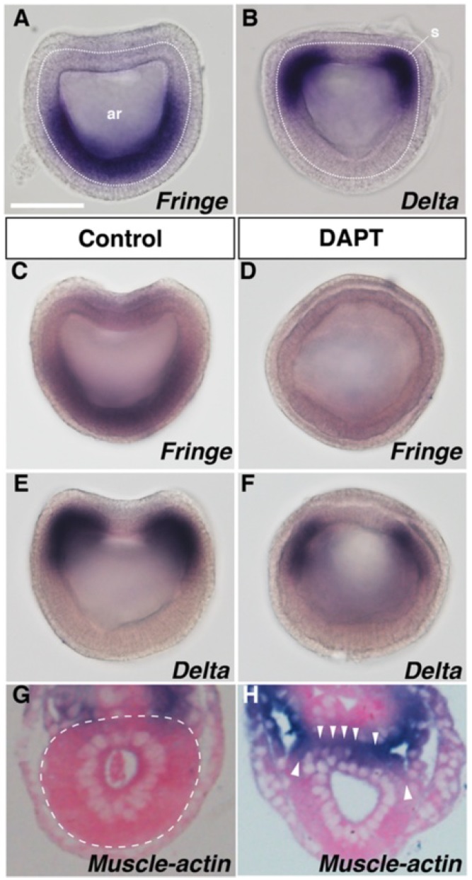
Fig. 2 Notch signalling controls the pinching off process of the
rostral somites. a–b BfFringe expressed in the anterior endoderm
(ventral part of the archenteron) and BfDelta was expressed in the
presumptive rostral somites at the late gastrula stage. Blastopore
views with the dorsal side up. The white dotted circle indicates the
archenteron. s somite, ar archenteron. Scale bar, 50 μm. c–f Effect of
100 μM DAPT treatment on BfFringe (n = 7, 100 %) or BfDelta (n = 8,
100 %). Anterior views with the dorsal side up. g In the DMSO-treated
control larval embryo, the segmental boundary between the somite
and the dorsal gut roof was clear (n = 1). h In larval embryos treated
with DAPT, the boundary was unclear and ectopic expression of
Muscle-actin was observed (n = 1). Transverse sections with the
dorsal side up. White arrowheads indicate somite and gut fusion
locations. The white dotted circle indicates the border between
the gut and somites Image published in: Onai T et al. (2015) © Onai et al. This image is reproduced with permission of the journal and the copyright holder. This is an open-access article distributed under the terms of the Creative Commons Attribution license Larger Image Printer Friendly View |