XB-IMG-150107
Xenbase Image ID: 150107

|
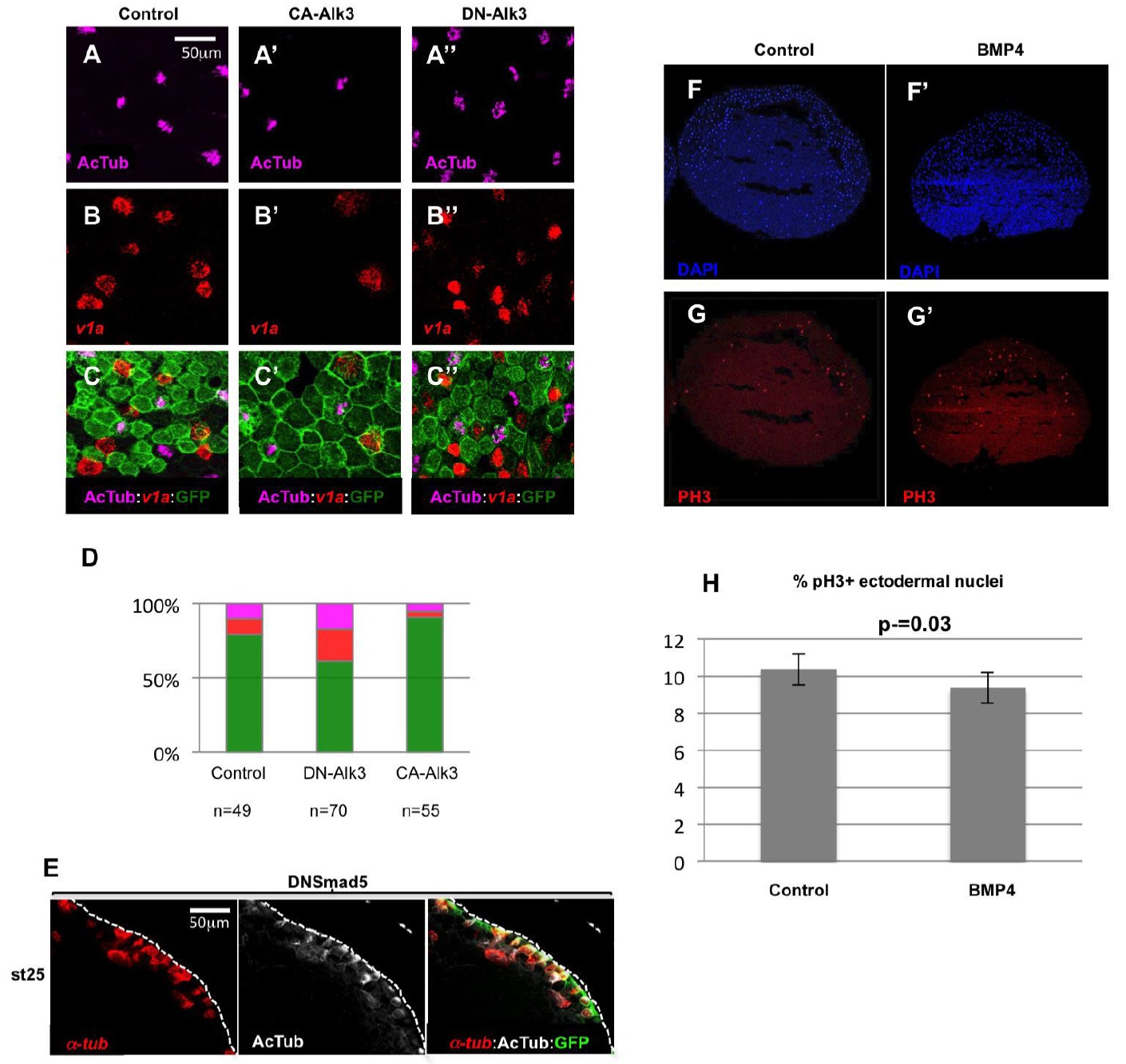
Figure S2: Interfering with the BMP pathway alters the numbers of MCCs and ionocytes in the developing Xenopus epidermis.
(A-D): 8-cell stage embryos were injected into the animal ventral blastomeres with synthetic mRNAs coding for GFP, alone (control; A, B, C) or together with mRNAs coding for a constitutively active (CA-Alk3; A', B', C') or a dominant negative (DN-Alk3; A'', B'', C'') version of the BMP receptor ALK3, then hybridized at stage 25 with a probe against the ionocyte marker v1a and antibodies against acetylated tubulin and against GFP. Activation of the BMP pathway by CA-Alk3 injection resulted in a decrease in the numbers of MCCs and Development | Supplementary Material
Development 142: doi:10.1242/dev.118679: Supplementary Material
ionocytes. Inhibition of the BMP pathway by DN-Alk3 injection results in an increase in the numbers of MCCs and ionocytes. (D): quantification of the different cellular populations in injected epidermal clones. The bars represent the total number of GFP positive cells scored. Magenta: acetylated tubulin positive MCCs; red: v1a positive ionocytes; green: GFP positive cells negative for both acetylated tubulin and v1a. (E): Embryos injected with a synthetic mRNA coding for a dominant negative form of the zebrafish BMP pathway nuclear effector Smad5 (dnSmad5), showed supernumerary ï¡-tubulin positive MCC precursors, which only partially managed to intercalate. (F-H): Embryos were injected in the blastocoele with BSA (control) or recombinant BMP4 protein (BMP4), fixed at stage 10, cryosectioned and immunostained with an antibody against phosphorylated histone H3 (red), a hallmark of cells in mitosis. DAPI (blue) was used to stain the nuclei. The graph in (H) shows the ratio of phospho-H3 positive nuclei to the total number of ectodermal DAPI stained nuclei. BMP4 injection did not significantly modify the number of mitotic nuclei compared to controls (Student's t-test). In F-G', the animal pole (ectoderm) is up. Image published in: Cibois M et al. (2015) Copyright © 2015. Image reproduced with permission of the Publisher.
Image source: Published Larger Image Printer Friendly View |
