XB-IMG-151249
Xenbase Image ID: 151249

|
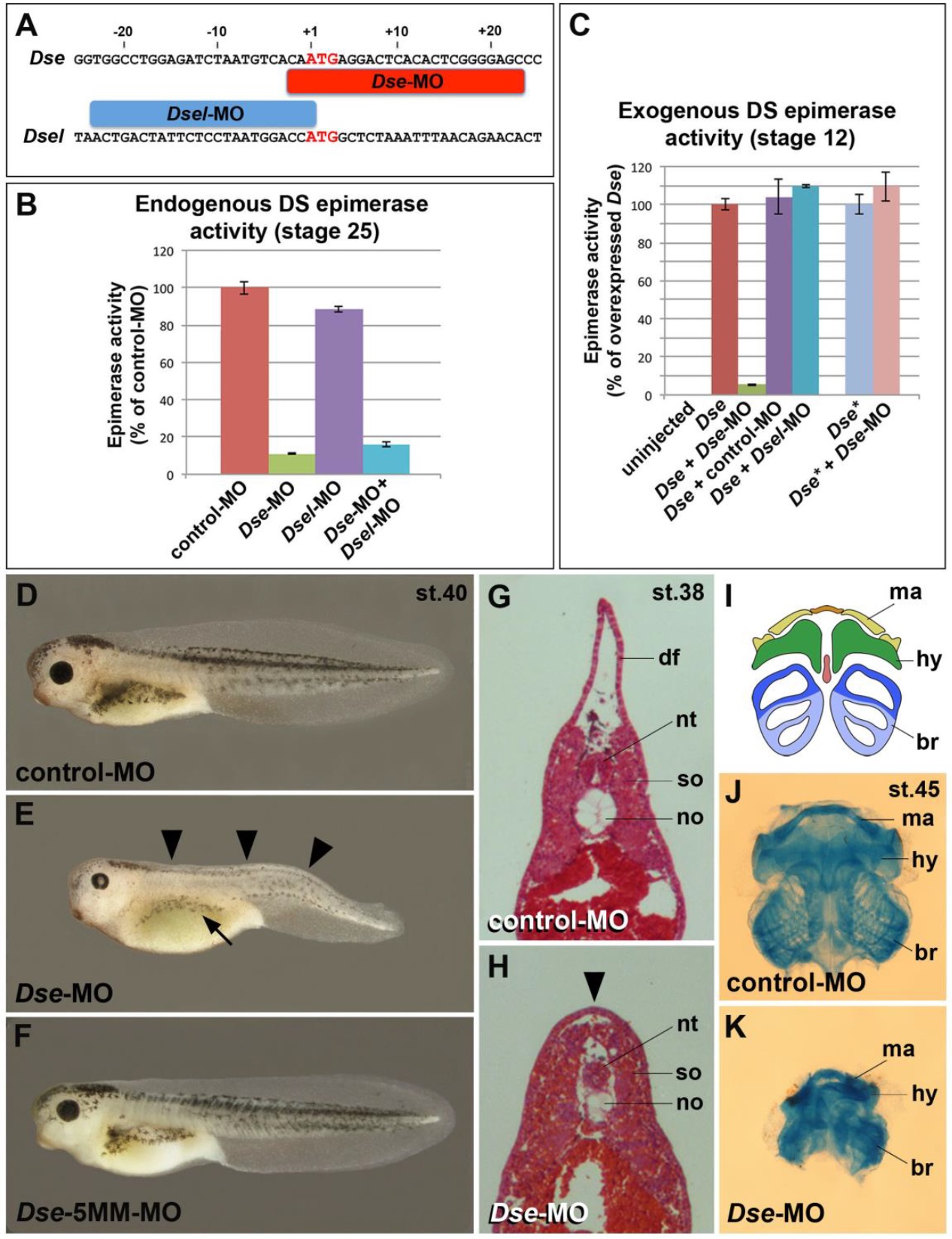
Figure 2. Knockdown of DS-epi1 reduces dermatan sulfate epimerase activity and neural crest-derived structures. (A) Morpholino oligonucleotides target the translation initiation sites of Dse and Dsel. (B) Endogenous DS epimerase activity is substantially decreased by Dse-MO and only little by Dsel-MO in stage 25 embryos. (C) Epimerase activity induced by the injection of 1 ng Dse mRNA is blocked by Dse-MO, but not by control-MO and Dsel-MO. The activity of 1 ng non-targeted Dse* mRNA is not affected by Dse-MO.
(D) Tadpole at stage 40 injected with control-MO. (E,F) Microinjection of Dse-MO, but not Dse-5MM-MO, induces small eyes, a lack of dorsal fin (arrowheads), and reduced melanocyte formation (arrow). (G,H) Transversal trunk sections of stage 38 embryos following hematoxylin-eosin staining. Note the lack of a dorsal fin (arrowhead), dorsally approaching somites and hypoplastic notochord in the Dse-morphant embryo. (I-K) Ventral view of head skeletons at stage 45 in a schematic overview (I) and following alcian blue staining (J,K). Injection of Dse-MO, but not control-MO, causes a reduction of NC-derived cartilage structures. br, branchial segment; hy, hyoid segment; df, dorsal fin; ma, mandibular segment; no, notochord; nt, neural tube; so, somite. Indicated phenotypes are shown as follows: D,
70/70; E, 71/114 (microcephaly), 92/114 (reduced dorsal fin), 70/114 (less
melanocytes); F, 63/63; G, 4/4; H, 4/4; J, 25/25; and K, 20/20. Image published in: Gouignard N et al. (2016) © 2016. Creative Commons Attribution license Larger Image Printer Friendly View |
