XB-IMG-151644
Xenbase Image ID: 151644
|
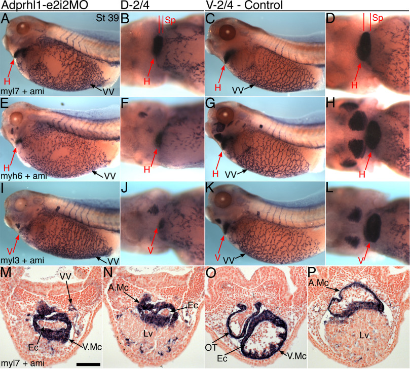
Fig. 2. Adprhl1 morpholino produces small, inert cardiac ventricles.A, B, E, F, I, J: Stage 39 tadpoles injected with Adprhl1-e2i2MO into d-2/4 blastomeres show detectable expression of
myl7 (mlc2, A, B),
myh6 (mhcα, E, F) and
myl3 (mlc1v, I, J)
in their smaller cardiac ventricles. C, D, G, H, K, L: Control stage 39 tadpoles that received the same morpholino into V-2/4 blastomeres. Left lateral views (A, C, E, G, I, K) and ventral views of heart (B, D, F, H, J, L), anterior to left. Vascular expression of ami (adipsin) is additionally shown to demonstrate that lateral vitelline vasculature, posterior cardinal vein and intersomitic vessels form in the absence of a functional heart beat (bare VV patches in A, E due to damage to tadpole during procedure). M, N: Representative transverse heart sections of the d-2/4-morpholino tadpole presented (A, B). O, P: Heart sections through the control tadpole (C, D). Section planes (Sp) indicated (B, D). Scale bar = 100 μm. H, heart; V, ventricle; A, atrium; Mc, myocardium; Ec, endocardium; OT, outflow tract; VV, vitelline vessel; Lv, liver. Image published in: Smith SJ et al. (2016) © 2016 The Authors. This image is reproduced with permission of the journal and the copyright holder. This is an open-access article distributed under the terms of the Creative Commons Attribution license
Image source: Published Larger Image Printer Friendly View |